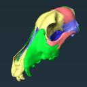
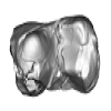
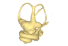
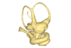
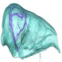
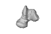
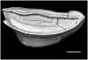
Explodable 3D Dog Skull for Veterinary Education
3D models of Ocnotherium skull
3D models of Kalakocetus, the earliest Cetacea
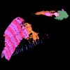
3D GM dataset of bird skeletal variation
Skeletal embryonic development in the catshark
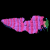
Bony connexions of the petrosal bone of extant hippos
bony labyrinth (14) , inner ear (11) , geometric morphometrics (10) , CT-scan (10) , Eocene (10) , Micro-CT (9) , Miocene (8)
Lionel Hautier (24) , Maëva Judith Orliac (23) , Laurent Marivaux (18) , Renaud Lebrun (14) , Rodolphe Tabuce (14) , Pierre-Olivier Antoine (13) , Bastien Mennecart (13)
|
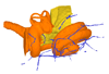
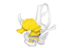
3D Printing an Explodable Dog Skull for Veterinary EducationWilliam C. Hooker
Published online: 17/12/2025 |

|
M3#1858PLYs of the segmented cranial bones with pre-fabricated magnetic casings and shelves for assembly following 3D printing Type: "3D_surfaces"doi: 10.18563/m3.sf.1858 state:published |
Download 3D surface file |
|
M3#1859PLYs of the segmented cranial bones of the "BOTTOM" cranial component. Downloadable for additional learning opportunities for students Type: "3D_surfaces"doi: 10.18563/m3.sf.1859 state:published |
Download 3D surface file |
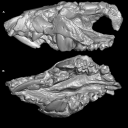
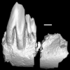
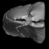
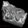
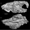
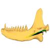
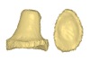
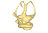
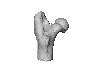
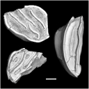
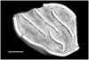
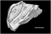
The present 3D Dataset contains the 3D models of the holotype and only specimen of Kalakocetus aurorae, a new cetacean retrieved from the Kalakot area in northwestern India. This specimen consists in a left hemimandible preserving the root of i3, p2, p4, m1 and m3 in situ. Its primitive morphology, with a tricuspid m3 morphologically intermediate between Raoellidae and Pakicetidae, makes it the first offshoot of Cetacea and provides crucial new elements to understand the setting up of the peculiar dental morphology of early cetaceans.
Kalakocetus aurorae GU/RJ/07 View specimen
|
M3#1803left hemi mandible with p2, p4, m1, m3 Type: "3D_surfaces"doi: 10.18563/m3.sf.1803 state:in_press |
Download 3D surface file |
|
M3#1804digitaly restored m1 Type: "3D_surfaces"doi: 10.18563/m3.sf.1804 state:in_press |
Download 3D surface file |
|
M3#1805digital restoration of complete mandible Type: "3D_surfaces"doi: 10.18563/m3.sf.1805 state:in_press |
Download 3D surface file |
|
M3#1810Scan Type: "3D_CT"doi: 10.18563/m3.sf.1810 state:in_press |
Download CT data |
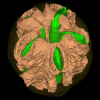
The present 3D Dataset contains the 3D models of two endocarps from the Oligocene of Baraval Quercy locality. These endocarps document new fossil genera within the Anacardiaceae family and illustrate the morphological diversity of this family during the Palaeogene. The CT-scan data were processed with ImageJ and Mimics Innovation Suite version 1.13 to reconstruct the specimens. Here we provide .stl files easy to read with the software Meshlab.
Palaeochoerospondias sheppeyensis UM-BAV-675 View specimen
|
M3#1895Surfaces of the endocarp, seeds and lacunae Type: "3D_surfaces"doi: 10.18563/m3.sf.1895 state:in_press |
Download 3D surface file |
Baravalosphaera operculata UM-BAV-689 View specimen
|
M3#1896Surfaces of the endocarp Type: "3D_surfaces"doi: 10.18563/m3.sf.1896 state:in_press |
Download 3D surface file |
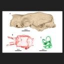
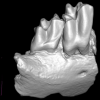
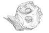
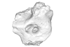
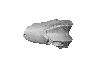
This contribution presents the three-dimensional digital models (i.e., skull, endocast, and inner ear) of a uniquely well-preserved and nearly complete skull (MCL 4228) attributed to the Late Pleistocene giant mylodontid ground sloth Ocnotherium giganteum, discovered in the Toca dos Ossos cave (Bahia State, Brazil). This specimen was described and figured in the following publication: Pujos et al. 2026: The neotropical giant ground sloth Ocnotherium giganteum (Xenarthra, Mylodontinae) from the Late Pleistocene of Brazil: anatomy, paleoneurology, and phylogenetic relationships. Zoological Journal of the Linnean Society. https://doi.org/10.1093/zoolinnean/zlag008
Ocnotherium giganteum MCL 4228 View specimen

|
M3#1870skull, endocast, inner ear Type: "3D_surfaces"doi: 10.18563/m3.sf.1870 state:in_press |
Download 3D surface file |
This contribution contains the 3D digital models of some fossil specimens of Wamradolops telloi Stutz and Pozodolops manuelorum Stutz (Metatheria: Polydolopimorphia), from several Palaeogene locations of Peruvian Amazonia. These taxa were described and analyzed in detail in the following publication: Stutz et al. (2026), Hidden diversity of Palaeogene metatherians: a new family of polydolopimorphian marsupials from Peruvian Amazonia. Zoological Journal of the Linnean Society. https://doi.org/10.1093/zoolinnean/zlag006.
Pozodolops manuelorum MUSM 4029 View specimen
|
M3#1874Pozodolops manuelorum, fragmentary left dentary with p3–m1 Type: "3D_surfaces"doi: 10.18563/m3.sf.1874 state:in_press |
Download 3D surface file |
Wamradolops telloi MUSM 4032 View specimen
|
M3#1875Wamradolops telloi, fragmentary right dentary with m1–m2 Type: "3D_surfaces"doi: 10.18563/m3.sf.1875 state:in_press |
Download 3D surface file |
Pozodolops manuelorum MUSM 4036 View specimen
|
M3#1876Pozodolops manuelorum, holotype, fragmentary right maxilla with M1 Type: "3D_surfaces"doi: 10.18563/m3.sf.1876 state:in_press |
Download 3D surface file |
Pozodolops manuelorum MUSM 4041 View specimen
|
M3#1877Pozodolops manuelorum, fragmentary right dentary with m1 Type: "3D_surfaces"doi: 10.18563/m3.sf.1877 state:in_press |
Download 3D surface file |
Pozodolops manuelorum MUSM 4058 View specimen
|
M3#1878Pozodolops manuelorum, fragmentary left P3 Type: "3D_surfaces"doi: 10.18563/m3.sf.1878 state:in_press |
Download 3D surface file |
Wamradolops telloi MUSM 4144 View specimen
|
M3#1879Wamradolops telloi, fragmentary left dentary with p2–m1 Type: "3D_surfaces"doi: 10.18563/m3.sf.1879 state:in_press |
Download 3D surface file |
Wamradolops telloi MUSM 4179 View specimen
|
M3#1880Wamradolops telloi, holotype, partial skull, with right P2–M4 and left I4–M3, plus boneless lower teeth below upper teeth, belonging to the same individual Type: "3D_surfaces"doi: 10.18563/m3.sf.1880 state:in_press |
Download 3D surface file |
Wamradolops telloi MUSM 4221 View specimen
|
M3#1881Wamradolops telloi, fragmentary of right dentary with p3–m2 Type: "3D_surfaces"doi: 10.18563/m3.sf.1881 state:in_press |
Download 3D surface file |
The present dataset contains 3D models used for illustration purposes in Schultz, J. A., Weaver, L. N., Jäger, K. R. K. & Grossnickle, D. M., 2026. Reexamining the evolutionary history of the mammalian medial pterygoid muscle. Evolution. The dataset includes 3D models based on micro-computed tomography (µCT) data of the postdentary area of the morganucodontan Morganucodon, the docodontan Docodon, the eutriconodontan Priacodon and the cladotherian Dryolestes. In addition, the dataset includes manually reconstructed schematic 3D models of the middle ear bones for the morganucodontan Morganucodon, the docodontan Docodon, the eutriconodontan Priacodon and schematic middle ear bones and a virtually rendered juvenile lower jaw of a juvenile monotreme Ornithorhynchus (based on illustrations of Zeller [1989]).
Morganucodon sp. NHM M84028 View specimen
|
M3#1883Lower jaw with manually reconstructed Meckel’s cartilage and schematic middle ear bones (Ectotympanic, Articular/part of Malleus, Surangular/ part of Malleus) Type: "3D_surfaces"doi: 10.18563/m3.sf.1883 state:in_press |
Download 3D surface file |
Priacodon fruitaensis LACM 120451 View specimen
|
M3#1884Lower jaw with manually reconstructed Meckel’s cartilage and schematic middle ear bones (Ectotympanic, Articular/part of Malleus, Surangular/part of Malleus) Type: "3D_surfaces"doi: 10.18563/m3.sf.1884 state:in_press |
Download 3D surface file |
Dryolestes leiriensis GuiMam 3-78 View specimen
|
M3#1888Lower jaw with manually reconstructed schematic Meckel’s cartilage Type: "3D_surfaces"doi: 10.18563/m3.sf.1888 state:in_press |
Download 3D surface file |
Docodon victor YPM10649, YPM13735, YPM11823 and YPM11826 View specimen
|
M3#1886Lower jaw with manually reconstructed Meckel’s cartilage and schematic middle ear bones (Ectotympanic, Articular/part of Malleus, Surangular/part of Malleus), for specification of jaw see Schultz et al. (2019) Type: "3D_surfaces"doi: 10.18563/m3.sf.1886 state:in_press |
Download 3D surface file |
Ornithorhynchus anatinus S32167 View specimen
|
M3#1887Lower jaw with manually reconstructed Meckel’s cartilage and schematic middle ear bones (Prearticular, Articular/part of the Malleus, Surangular/part of Malleus, Angular/Incus) Type: "3D_surfaces"doi: 10.18563/m3.sf.1887 state:in_press |
Download 3D surface file |

The present 3D Dataset contains the 3D models of the two papionine remains found near Gabes and analyzed in Ksila et al. 2026 “A continental Messinian vertebrate fauna from the Ouedhref area, Southeast Tunisia.”
Macaca sp. UTM-O-Sa60 View specimen
|
M3#1872Right m1 or m2 Type: "3D_surfaces"doi: 10.18563/m3.sf.1872 state:in_press |
Download 3D surface file |
Macaca sp. UTM-O-Br6 View specimen
|
M3#1871Left upper canine Type: "3D_surfaces"doi: 10.18563/m3.sf.1871 state:in_press |
Download 3D surface file |
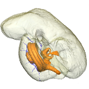
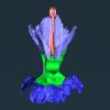
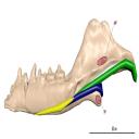
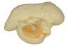
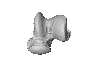
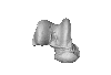
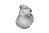
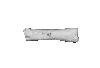
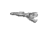
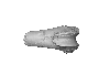
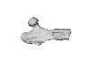
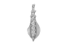
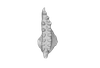
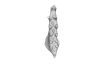
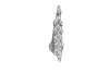
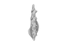
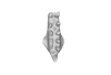
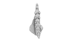
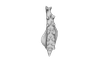
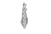
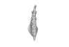
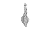
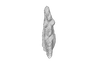
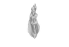
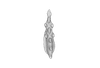
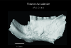
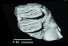
In this contribution, we describe the external and internal morphology of a delphinid petrosal bone collected from Ahu Tahai, a burial site located on the Southwestern coast of Easter Island, at Hangaroa. We discuss the taxonomic attribution of this archaeological item and describe its internal structures based on µCT data, including the bony labyrinth and the nerve and vein patterns. Identification of the nerves exists lead us to relocate the identification of the foramen singulare in delphinid petrosals.
indet. indet. AT1 View specimen
|
M3#420Stapes Type: "3D_surfaces"doi: 10.18563/m3.sf.420 state:published |
Download 3D surface file |
|
M3#421petrosal bone Type: "3D_surfaces"doi: 10.18563/m3.sf.421 state:published |
Download 3D surface file |
|
M3#422in situ bony labyrinth Type: "3D_surfaces"doi: 10.18563/m3.sf.422 state:published |
Download 3D surface file |
|
M3#423bony labyrinth and associated nerves and blood vessels Type: "3D_surfaces"doi: 10.18563/m3.sf.423 state:published |
Download 3D surface file |
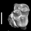
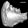
This contribution contains the 3D model of the holotype of Chambius kasserinensis, the basalmost ‘elephant-shrew’ figured in the following publication: New remains of Chambius kasserinensis from the Eocene of Tunisia and evaluation of proposed affinities for Macroscelidea (Mammalia, Afrotheria). https://doi.org/10.1080/08912963.2017.1297433
Chambius kasserinensis CBI-1-06 View specimen
|
M3#1463D model of the holotype maxilla of Chambius kasserinensis. The 3D surface was extracted manually from the limestone matrix within AVIZO 9.2 Type: "3D_surfaces"doi: 10.18563/m3.sf.146 state:published |
Download 3D surface file |
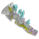
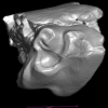
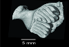
This contribution contains the 3D models described and figured in the publication entitled "The petrosal and bony labyrinth of Diplobune minor, an enigmatic Artiodactyla from the Oligocene of Western Europe" by Orliac, Araújo, and Lihoreau published in Journal of Morphology (Orliac et al. 2017) https://doi.org/10.1002/jmor.20702.
Diplobune minor UM ITD 1079 View specimen
|
M3#138right bony labyrinth of Diplobune minor from Itardies, France Type: "3D_surfaces"doi: 10.18563/m3.sf.138 state:published |
Download 3D surface file |
|
M3#139right isolated petrosal of Diplobune minor from Itardies, France Type: "3D_surfaces"doi: 10.18563/m3.sf.139 state:published |
Download 3D surface file |
Diplobune minor UM ITD 1080 View specimen
|
M3#140left bony labyrinth of Diplobune minor from Itardies, France Type: "3D_surfaces"doi: 10.18563/m3.sf.140 state:published |
Download 3D surface file |
|
M3#141left isolated petrosal of Diplobune minor from Itardies, France Type: "3D_surfaces"doi: 10.18563/m3.sf.141 state:published |
Download 3D surface file |
Diplobune minor UM ITD 1081 View specimen
|
M3#142right bony labyrinth and associated nerves and veins of Diplobune minor from Itardies, France Type: "3D_surfaces"doi: 10.18563/m3.sf.142 state:published |
Download 3D surface file |
|
M3#143right isolated petrosal of Diplobune minor from Itardies, France Type: "3D_surfaces"doi: 10.18563/m3.sf.143 state:published |
Download 3D surface file |
Diplobune minor UM ITD 1083 View specimen
|
M3#144left bony labyrinth of Diplobune minor from Itardies, France Type: "3D_surfaces"doi: 10.18563/m3.sf.144 state:published |
Download 3D surface file |
|
M3#145left petrosal of Diplobune minor from Itardies, France Type: "3D_surfaces"doi: 10.18563/m3.sf.145 state:published |
Download 3D surface file |
The present 3D Dataset contains a selection of 3D models analyzed in Billet G, Hautier L, Gaudin TJ, Flynn JJ, Ruf I, Carrillo JD, Ladevèze S, Lehmann T, Nicolas V, Orliac MJ, Tornero C, Wible JR, Wong N, Gaubert P. Submitted. Brain drain: Exceptional pattern of calvarial venation in pangolins and its phylogenetic significance for Ferae. Zoological Journal of the Linnean Society.
Phataginus tricuspis NHM-UK 48.13.26 View specimen

|
M3#1847cranium & intradiploic canals (sinuses & diploic veins) Type: "3D_surfaces"doi: 10.18563/m3.sf.1847 state:in_press |
Download 3D surface file |
Manis javanica NHM-UK 9.1.5.858 View specimen

|
M3#1848cranium & intradiploic canals (sinuses & diploic veins) Type: "3D_surfaces"doi: 10.18563/m3.sf.1848 state:in_press |
Download 3D surface file |
Felis silvestris UM-ZOOL-149N View specimen

|
M3#1849cranium & intradiploic canals (sinuses & diploic veins) Type: "3D_surfaces"doi: 10.18563/m3.sf.1849 state:in_press |
Download 3D surface file |
Erinaceus europaeus SMNS40759 View specimen

|
M3#1850cranium & intradiploic canals (sinuses & diploic veins) Type: "3D_surfaces"doi: 10.18563/m3.sf.1850 state:in_press |
Download 3D surface file |
Pterodon dasyuroides MNHN.F.Qu8301 View specimen

|
M3#1851cranium & intradiploic canals (sinuses & diploic veins) Type: "3D_surfaces"doi: 10.18563/m3.sf.1851 state:in_press |
Download 3D surface file |
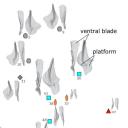
The present 3D dataset contains 15 specimens selected from the 69 3D models analyzed in the paper “3D topography as an indicator of change in food processing ability in the conodont genus Palmatolepis elements”. 3D topographic analysis of Palmatolepis P1 conodont elements from the Late Devonian period revealed an increase in blade sharpness together with a reduction in platform size. This indicates morphofunctional adaptation to more efficient prey processing.
Palmatolepis manticolepis UM CTB 144 View specimen

|
M3#1814Palmatolepis Manticolepis Type: "3D_surfaces"doi: 10.18563/m3.sf.1814 state:in_press |
Download 3D surface file |
Palmatolepis manticolepis UM CTB 151 View specimen

|
M3#1815Palmatolepis Manticolepis Type: "3D_surfaces"doi: 10.18563/m3.sf.1815 state:in_press |
Download 3D surface file |
Palmatolepis manticolepis UM CTB 078 View specimen

|
M3#1816Palmatolepis Manticolepis Type: "3D_surfaces"doi: 10.18563/m3.sf.1816 state:in_press |
Download 3D surface file |
Palmatolepis rhomboidea UM CTB 172 View specimen

|
M3#1817Palmatolepis rhomboidea Type: "3D_surfaces"doi: 10.18563/m3.sf.1817 state:in_press |
Download 3D surface file |
Palmatolepis glabra UM CTB 080 View specimen

|
M3#1818Palmatolepis glabra Type: "3D_surfaces"doi: 10.18563/m3.sf.1818 state:in_press |
Download 3D surface file |
Palmatolepis glabra UM CTB 177 View specimen

|
M3#1819Palmatolepis glabra Type: "3D_surfaces"doi: 10.18563/m3.sf.1819 state:in_press |
Download 3D surface file |
Palmatolepis glabra UM CTB 178 View specimen

|
M3#1820Palmatolepis glabra Type: "3D_surfaces"doi: 10.18563/m3.sf.1820 state:in_press |
Download 3D surface file |
Palmatolepis glabra UM CTB 179 View specimen

|
M3#1821Palmatolepis glabra Type: "3D_surfaces"doi: 10.18563/m3.sf.1821 state:in_press |
Download 3D surface file |
Palmatolepis gracilis UM CTB 186 View specimen

|
M3#1822Palmatolepis gracilis Type: "3D_surfaces"doi: 10.18563/m3.sf.1822 state:in_press |
Download 3D surface file |
Palmatolepis perlobata UM CTB 187 View specimen

|
M3#1823Palmatolepis perlobata Type: "3D_surfaces"doi: 10.18563/m3.sf.1823 state:in_press |
Download 3D surface file |
Palmatolepis perlobata UM CTB 189 View specimen

|
M3#1824Palmatolepis perlobata Type: "3D_surfaces"doi: 10.18563/m3.sf.1824 state:in_press |
Download 3D surface file |
Palmatolepis gracilis UM CTB 190 View specimen

|
M3#1825Palmatolepis gracilis Type: "3D_surfaces"doi: 10.18563/m3.sf.1825 state:in_press |
Download 3D surface file |
Palmatolepis perlobata UM CTB 191 View specimen

|
M3#1826Palmatolepis perlobata Type: "3D_surfaces"doi: 10.18563/m3.sf.1826 state:in_press |
Download 3D surface file |
Palmatolepis gracilis UM CTB 197 View specimen

|
M3#1827Palmatolepis gracilis Type: "3D_surfaces"doi: 10.18563/m3.sf.1827 state:in_press |
Download 3D surface file |
Palmatolepis gracilis UM CTB 200 View specimen

|
M3#1828Palmatolepis gracilis Type: "3D_surfaces"doi: 10.18563/m3.sf.1828 state:in_press |
Download 3D surface file |
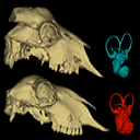
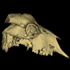
This contribution contains the 3D models described and figured in the following publication:Skull and Inner Ear Morphometrics in Sheep and Goats: Species and Breed Differentiation with Bioarchaeological Applications (Hemelsdael et al. submitted). The models include the external surface of a complete skull and inner ear of both a sheep (Ovis aries) and a goat (Capra hircus), generated from micro-CT scans. In the associated paper, we used 3D geometric morphometric data to assess inter and intra (i.e. between breeds) discrimination based on complete skulls, skull fragments and the semi-circular canals of the inner ear.
Capra hircus Amp_1 View specimen
|
M3#1806Skull of the goat Amp_1 Type: "3D_surfaces"doi: 10.18563/m3.sf.1806 state:published |
Download 3D surface file |
|
M3#1807Inner ear of the goat Amp_1 Type: "3D_surfaces"doi: 10.18563/m3.sf.1807 state:published |
Download 3D surface file |
Ovis aries UM_RR_2331 View specimen
|
M3#1808Skull of the sheep UM_RR_2331 Type: "3D_surfaces"doi: 10.18563/m3.sf.1808 state:published |
Download 3D surface file |
|
M3#1809Inner ear of the sheep UM_RR_2331 Type: "3D_surfaces"doi: 10.18563/m3.sf.1809 state:published |
Download 3D surface file |
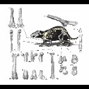
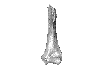
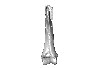
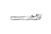
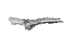
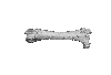
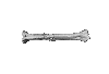
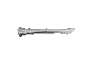
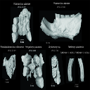
This contribution contains the 3D models of postcranial bones (humerus, ulna, innominate, femur, tibia, astragalus, navicular, and metatarsal III) described and figured in the following publication: “Postcranial morphology of the extinct rodent Neoepiblema (Rodentia: Chinchilloidea): insights into the paleobiology of neoepiblemids”.
Neoepiblema acreensis UFAC 3549 View specimen
|
M3#719UFAC 3549, left humerus missing the proximal region. Type: "3D_surfaces"doi: 10.18563/m3.sf.719 state:published |
Download 3D surface file |
Neoepiblema acreensis UFAC 5076 View specimen
|
M3#720UFAC 5076, right humerus missing the proximal region. Type: "3D_surfaces"doi: 10.18563/m3.sf.720 state:published |
Download 3D surface file |
Neoepiblema acreensis UFAC 1939 View specimen
|
M3#721UFAC 1939, right ulna missing the olecranon epiphysis and the distal region. Type: "3D_surfaces"doi: 10.18563/m3.sf.721 state:published |
Download 3D surface file |
Neoepiblema acreensis UFAC 3697 View specimen
|
M3#722UFAC 3697, right innominate bone. Type: "3D_surfaces"doi: 10.18563/m3.sf.722 state:published |
Download 3D surface file |
Neoepiblema acreensis UFAC 2574 View specimen
|
M3#723UFAC 2574, proximal region of a left femur. Type: "3D_surfaces"doi: 10.18563/m3.sf.723 state:published |
Download 3D surface file |
Neoepiblema acreensis UFAC 2937 View specimen
|
M3#724UFAC 2937, right femur with damaged proximal region. Type: "3D_surfaces"doi: 10.18563/m3.sf.724 state:published |
Download 3D surface file |
Neoepiblema acreensis UFAC 2210 View specimen
|
M3#725UFAC 2210, distal region of a right femur. Type: "3D_surfaces"doi: 10.18563/m3.sf.725 state:published |
Download 3D surface file |
Neoepiblema acreensis UFAC 1887 View specimen
|
M3#726UFAC 1887, right tibia Type: "3D_surfaces"doi: 10.18563/m3.sf.726 state:published |
Download 3D surface file |
Neoepiblema acreensis UFAC 1840 View specimen
|
M3#727UFAC 1840, left astragalus. Type: "3D_surfaces"doi: 10.18563/m3.sf.727 state:published |
Download 3D surface file |
Neoepiblema acreensis UFAC 2549 View specimen
|
M3#728UFAC 2549, right astragalus. Type: "3D_surfaces"doi: 10.18563/m3.sf.728 state:published |
Download 3D surface file |
Neoepiblema acreensis UFAC 3672 View specimen
|
M3#729UFAC 3672, right navicular. Type: "3D_surfaces"doi: 10.18563/m3.sf.729 state:published |
Download 3D surface file |
Neoepiblema acreensis UFAC 2116 View specimen
|
M3#730UFAC 2116, left metatarsal III. Type: "3D_surfaces"doi: 10.18563/m3.sf.730 state:published |
Download 3D surface file |
Neoepiblema horridula UFAC 3260 View specimen
|
M3#731UFAC 3260, fragmented left innominate. Type: "3D_surfaces"doi: 10.18563/m3.sf.731 state:published |
Download 3D surface file |
Neoepiblema horridula UFAC 2620 View specimen
|
M3#732UFAC 2620, distal region of a right femur. Type: "3D_surfaces"doi: 10.18563/m3.sf.732 state:published |
Download 3D surface file |
Neoepiblema horridula UFAC 2737 View specimen
|
M3#733UFAC 2737, proximal region of right femur. Type: "3D_surfaces"doi: 10.18563/m3.sf.733 state:published |
Download 3D surface file |
Neoepiblema horridula UFAC 3202 View specimen
|
M3#734UFAC 3202, right tibia, missing the proximalmost and distal portions. Type: "3D_surfaces"doi: 10.18563/m3.sf.734 state:published |
Download 3D surface file |
Neoepiblema horridula UFAC 3212 View specimen
|
M3#735UFAC 3212, left astragalus. Type: "3D_surfaces"doi: 10.18563/m3.sf.735 state:published |
Download 3D surface file |
This contribution contains the 3D models of the fossil teeth of two chinchilloid caviomorph rodents (Borikenomys praecursor and Chinchilloidea gen. et sp. indet.) discovered from lower Oligocene deposits of Puerto Rico, San Sebastian Formation (locality LACM Loc. 8060). These fossils were described and figured in the following publication: Marivaux et al. (2020), Early Oligocene chinchilloid caviomorphs from Puerto Rico and the initial rodent colonization of the West Indies. Proceedings of the Royal Society B. http://dx.doi.org/10.1098/rspb.2019.2806
Borikenomys praecursor LACM 162447 View specimen
|
M3#638Right lower m3. This isolated tooth was scanned with a resolution of 6 µm using a μ-CT-scanning station EasyTom 150 / Rx Solutions (Montpellier RIO Imaging, ISE-M, Montpellier, France). AVIZO 7.1 (Visualization Sciences Group) software was used for visualization, segmentation, and 3D rendering. The specimen was prepared within a “labelfield” module of AVIZO, using the segmentation threshold selection tool. Type: "3D_surfaces"doi: 10.18563/m3.sf.638 state:published |
Download 3D surface file |
Borikenomys praecursor LACM 162446 View specimen
|
M3#639Fragment of lower molar (most of the mesial part). This isolated broken tooth was scanned with a resolution of 6 µm using a μ-CT-scanning station EasyTom 150 / Rx Solutions (Montpellier RIO Imaging, ISE-M, Montpellier, France). AVIZO 7.1 (Visualization Sciences Group) software was used for visualization, segmentation, and 3D rendering. The specimen was prepared within a “labelfield” module of AVIZO, using the segmentation threshold selection tool. Type: "3D_surfaces"doi: 10.18563/m3.sf.639 state:published |
Download 3D surface file |
indet indet LACM 162448 View specimen
|
M3#640Fragment of either an upper tooth (mesial laminae) or a lower tooth (distal laminae). The specimen was scanned with a resolution of 6 µm using a μ-CT-scanning station EasyTom 150 / Rx Solutions (Montpellier RIO Imaging, ISE-M, Montpellier, France). AVIZO 7.1 (Visualization Sciences Group) software was used for visualization, segmentation, and 3D rendering. This fragment of tooth was prepared within a “labelfield” module of AVIZO, using the segmentation threshold selection tool. Type: "3D_surfaces"doi: 10.18563/m3.sf.640 state:published |
Download 3D surface file |
This contribution contains the 3D model described and figured in the following publication: Albino, A., Carrillo-Briceño, J. D. & Neenan, J. M. 2016. An enigmatic aquatic snake from the Cenomanian of northern South America. PeerJ 4:e2027 http://dx.doi.org/10.7717/peerj.2027
Lunaophis aquaticus MCNC-1827-F View specimen
|
M3#116Articulated precloacal vertebrae of Lunaophis aquaticus Type: "3D_surfaces"doi: 10.18563/m3.sf.116 state:published |
Download 3D surface file |
The present 3D Dataset contains the 3D models analyzed in the publication: Head anatomy and phylogenomics show the Carboniferous giant Arthropleura was a relative to both millipedes and centipedes. Lhéritier Mickaël, Edgecombe Gregory D., Garwodd Russell J., Buisson Adrien, Gerbe Alexis, Mongiardino Koch Nicolás, Vannier Jean, Escarguel Gilles, Adrien Jérome, Fernandez Vincent, Bergeret-Medina Aude, Giupponi Alexandra and Perrier Vincent. Sciences Advances. https://www.science.org/doi/10.1126/sciadv.adp6362
Arthropleura sp. MNHN.F.SOT002123 View specimen
|
M3#1481Reconstitution of MNH.F.SOT002123 made from Phoenix X-ray Phoenix V|tome|x CT-scan Type: "3D_surfaces"doi: 10.18563/m3.sf.1481 state:published |
Download 3D surface file |
|
M3#1482Ct-scan (X-ray Phoenix V|tome|x) of MNHN.F.SOT002123 Type: "3D_CT"doi: 10.18563/m3.sf.1482 state:published |
Download CT data |
|
M3#1484Reconstitution of MNH.F.SOT002123 made from synchrotron X-ray micro-Computed tomography Type: "3D_surfaces"doi: 10.18563/m3.sf.1484 state:published |
Download 3D surface file |
|
M3#1485Synchrotron data of MNHN.F.SOT002123 (bin4) Type: "3D_CT"doi: 10.18563/m3.sf.1485 state:published |
Download CT data |
Arthropleura sp. MNHN.F.SOT002118 View specimen
|
M3#1480Reconstitution of MNH.F.SOT002118 made from Phoenix X-ray Phoenix V|tome|x CT-scan Type: "3D_surfaces"doi: 10.18563/m3.sf.1480 state:published |
Download 3D surface file |
|
M3#1483Ct-scan (X-ray Phoenix V|tome|x) of MNHN.F.SOT002118 Type: "3D_CT"doi: 10.18563/m3.sf.1483 state:published |
Download CT data |
Arthropleura sp. MNHN.F.SOT002123 (synchrotron data) View specimen
This contribution contains the 3D models of a set of Famennian conodont elements belonging to the species Icriodus alternatus analyzed in the following publication: Girard et al. 2022: Deciphering the morphological variation and its ontogenetic dynamics in the Late Devonian conodont Icriodus alternatus.
Icriodus alternatus UM BUS 031 View specimen
|
M3#887conodont element Type: "3D_surfaces"doi: 10.18563/m3.sf.887 state:published |
Download 3D surface file |
Icriodus alternatus UM BUS 032 View specimen
|
M3#888conodont element Type: "3D_surfaces"doi: 10.18563/m3.sf.888 state:published |
Download 3D surface file |
Icriodus alternatus UM BUS 033 View specimen
|
M3#889conodont element Type: "3D_surfaces"doi: 10.18563/m3.sf.889 state:published |
Download 3D surface file |
Icriodus alternatus UM BUS 034 View specimen
|
M3#890conodont element Type: "3D_surfaces"doi: 10.18563/m3.sf.890 state:published |
Download 3D surface file |
Icriodus alternatus UM BUS 035 View specimen
|
M3#891conodont element Type: "3D_surfaces"doi: 10.18563/m3.sf.891 state:published |
Download 3D surface file |
Icriodus alternatus UM BUS 036 View specimen
|
M3#892conodont element Type: "3D_surfaces"doi: 10.18563/m3.sf.892 state:published |
Download 3D surface file |
Icriodus alternatus UM BUS 037 View specimen
|
M3#893conodont element Type: "3D_surfaces"doi: 10.18563/m3.sf.893 state:published |
Download 3D surface file |
Icriodus alternatus UM BUS 038 View specimen
|
M3#894conodont element Type: "3D_surfaces"doi: 10.18563/m3.sf.894 state:published |
Download 3D surface file |
Icriodus alternatus UM BUS 039 View specimen
|
M3#895conodont element Type: "3D_surfaces"doi: 10.18563/m3.sf.895 state:published |
Download 3D surface file |
Icriodus alternatus UM BUS 040 View specimen
|
M3#896conodont element Type: "3D_surfaces"doi: 10.18563/m3.sf.896 state:published |
Download 3D surface file |
Icriodus alternatus UM BUS 041 View specimen
|
M3#897conodont element Type: "3D_surfaces"doi: 10.18563/m3.sf.897 state:published |
Download 3D surface file |
Icriodus alternatus UM BUS 042 View specimen
|
M3#898conodont element Type: "3D_surfaces"doi: 10.18563/m3.sf.898 state:published |
Download 3D surface file |
Icriodus alternatus UM BUS 043 View specimen
|
M3#899conodont element Type: "3D_surfaces"doi: 10.18563/m3.sf.899 state:published |
Download 3D surface file |
Icriodus alternatus UM BUS 044 View specimen
|
M3#900conodont element Type: "3D_surfaces"doi: 10.18563/m3.sf.900 state:published |
Download 3D surface file |
Icriodus alternatus UM BUS 045 View specimen
|
M3#901conodont element Type: "3D_surfaces"doi: 10.18563/m3.sf.901 state:published |
Download 3D surface file |
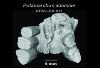
The presented dataset contains the 3D surface scan of the holotype of Birgeria americana, a partial skull described and depicted in: Romano, C., Jenks, J.F., Jattiot, R., Scheyer, T.M., Bylund, K.G. & Bucher, H. 2017. Marine Early Triassic Actinopterygii from Elko County (Nevada, USA): implications for the Smithian equatorial vertebrate eclipse. Journal of Paleontology. https://doi.org/10.1017/jpa.2017.36 .
Birgeria americana NMMNH P-66225 View specimen
|
M3#175NMMNH P-66225 is from upper lower Smithian to lower upper Smithian beds (Thaynes Group). The collecting site is located about 2.75 km south-southeast of the Winecup Ranch, east-central Elko County, Nevada, USA. P-66225 is a partial skull preserved within a large limestone nodule, with its right side exposed. It preserves the portion between the cleithrum posteriorly, and the level of the hind margin of the orbital opening anteriorly. The fossil has a length of 26 cm. Type: "3D_surfaces"doi: 10.18563/m3.sf.175 state:published |
Download 3D surface file |
This contribution contains 3D models of extinct rodents Dinomyidae from Miocene and Quaternary of Brazil. The Miocene specimens that were digitalized include the holotypes of Potamarchus adamiae, Pseudopotamarchus villanuevai, and Ferigolomys pacarana collected in the Solimões Formation (Upper Miocene), northern Brazil. The Quaternary specimens are the holotype and paratype of Niedemys piauiensis, found in Upper Pleistocene deposits from northeast Brazil.
Potamarchus adamiae UFAC-CS 011 View specimen
|
M3#410UFAC-CS 011 – holotype, palatal region of the skull with cheek teeth Type: "3D_surfaces"doi: 10.18563/m3.sf.410 state:published |
Download 3D surface file |
Potamarchus adamiae UFAC-CS 043 View specimen
|
M3#411UFAC-CS 043, left dentary with cheek teeth Type: "3D_surfaces"doi: 10.18563/m3.sf.411 state:published |
Download 3D surface file |
Pseudopotamarchus villanuevai UFAC 4762 View specimen
|
M3#412UFAC 4762 – holotype, incomplete right maxilla with cheek teeth Type: "3D_surfaces"doi: 10.18563/m3.sf.412 state:published |
Download 3D surface file |
Ferigolomys pacarana UFAC 6460 View specimen
|
M3#413UFAC 6460 – holotype, palatal region of the skull with cheek teeth Type: "3D_surfaces"doi: 10.18563/m3.sf.413 state:published |
Download 3D surface file |
Drytomomys sp. UFAC 2742 View specimen

|
M3#414UFAC 2742, right dentary with cheek teeth Type: "3D_surfaces"doi: 10.18563/m3.sf.414 state:published |
Download 3D surface file |
Niedemys piauiensis FUMDHAM 113-146365-2 View specimen
|
M3#418FUMDHAM 113-146365-2 - holotype, upper right tooth Type: "3D_surfaces"doi: 10.18563/m3.sf.418 state:published |
Download 3D surface file |
Niedemys piauiensis FUMDHAM 113-145304-2 View specimen
|
M3#419FUMDHAM 113-145304-2 - paratype, left lower molar Type: "3D_surfaces"doi: 10.18563/m3.sf.419 state:published |
Download 3D surface file |

