Explodable 3D Dog Skull for Veterinary Education
3D models of Ocnotherium skull
3D models of Kalakocetus, the earliest Cetacea
3D GM dataset of bird skeletal variation
Skeletal embryonic development in the catshark
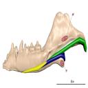
Bony connexions of the petrosal bone of extant hippos
bony labyrinth (14) , inner ear (11) , geometric morphometrics (10) , CT-scan (10) , Eocene (10) , Micro-CT (9) , Miocene (8)
Lionel Hautier (24) , Maëva Judith Orliac (23) , Laurent Marivaux (18) , Renaud Lebrun (14) , Rodolphe Tabuce (14) , Pierre-Olivier Antoine (13) , Bastien Mennecart (13)
MorphoMuseuM, also referred to as M3, is a peer reviewed, online data journal that publishes 3D models of vertebrates, including models of type specimens, anatomy atlases, reconstruction of deformed or damaged specimens, and 3D datasets (see https://doi.org/10.1017/scs.2017.14 for details).
M3 comes along with a free software, MorphoDig, which contains a set of tools for editing, positioning, deforming, labelling, measuring and rendering sets of 3D surfaces.
All 3D data presented on this website are licensed under a Creative Commons Attribution-NonCommercial 4.0. International License. 
|
3D models related to the publication: Fruits of Anacardiaceae from the Early Oligocene of Baraval Quercy locality, southwestern FranceMuratcan Ersoy, Yiyun Chen, Anaïs Boura
Published online: 13/05/2026 |
|
M3#1895Surfaces of the endocarp, seeds and lacunae Type: "3D_surfaces"doi: 10.18563/m3.sf.1895 state:in_press |
Download 3D surface file |
Baravalosphaera operculata UM-BAV-689 View specimen
|
M3#1896Surfaces of the endocarp Type: "3D_surfaces"doi: 10.18563/m3.sf.1896 state:in_press |
Download 3D surface file |
The present dataset contains 3D models used for illustration purposes in Schultz, J. A., Weaver, L. N., Jäger, K. R. K. & Grossnickle, D. M., 2026. Reexamining the evolutionary history of the mammalian medial pterygoid muscle. Evolution. The dataset includes 3D models based on micro-computed tomography (µCT) data of the postdentary area of the morganucodontan Morganucodon, the docodontan Docodon, the eutriconodontan Priacodon and the cladotherian Dryolestes. In addition, the dataset includes manually reconstructed schematic 3D models of the middle ear bones for the morganucodontan Morganucodon, the docodontan Docodon, the eutriconodontan Priacodon and schematic middle ear bones and a virtually rendered juvenile lower jaw of a juvenile monotreme Ornithorhynchus (based on illustrations of Zeller [1989]).
Morganucodon sp. NHM M84028 View specimen
|
M3#1883Lower jaw with manually reconstructed Meckel’s cartilage and schematic middle ear bones (Ectotympanic, Articular/part of Malleus, Surangular/ part of Malleus) Type: "3D_surfaces"doi: 10.18563/m3.sf.1883 state:in_press |
Download 3D surface file |
Priacodon fruitaensis LACM 120451 View specimen
|
M3#1884Lower jaw with manually reconstructed Meckel’s cartilage and schematic middle ear bones (Ectotympanic, Articular/part of Malleus, Surangular/part of Malleus) Type: "3D_surfaces"doi: 10.18563/m3.sf.1884 state:in_press |
Download 3D surface file |
Dryolestes leiriensis GuiMam 3-78 View specimen
|
M3#1888Lower jaw with manually reconstructed schematic Meckel’s cartilage Type: "3D_surfaces"doi: 10.18563/m3.sf.1888 state:in_press |
Download 3D surface file |
Docodon victor YPM10649, YPM13735, YPM11823 and YPM11826 View specimen
|
M3#1886Lower jaw with manually reconstructed Meckel’s cartilage and schematic middle ear bones (Ectotympanic, Articular/part of Malleus, Surangular/part of Malleus), for specification of jaw see Schultz et al. (2019) Type: "3D_surfaces"doi: 10.18563/m3.sf.1886 state:in_press |
Download 3D surface file |
Ornithorhynchus anatinus S32167 View specimen
|
M3#1887Lower jaw with manually reconstructed Meckel’s cartilage and schematic middle ear bones (Prearticular, Articular/part of the Malleus, Surangular/part of Malleus, Angular/Incus) Type: "3D_surfaces"doi: 10.18563/m3.sf.1887 state:in_press |
Download 3D surface file |
This contribution contains the 3D digital models of some fossil specimens of Wamradolops telloi Stutz and Pozodolops manuelorum Stutz (Metatheria: Polydolopimorphia), from several Palaeogene locations of Peruvian Amazonia. These taxa were described and analyzed in detail in the following publication: Stutz et al. (2026), Hidden diversity of Palaeogene metatherians: a new family of polydolopimorphian marsupials from Peruvian Amazonia. Zoological Journal of the Linnean Society. https://doi.org/10.1093/zoolinnean/zlag006.
Pozodolops manuelorum MUSM 4029 View specimen
|
M3#1874Pozodolops manuelorum, fragmentary left dentary with p3–m1 Type: "3D_surfaces"doi: 10.18563/m3.sf.1874 state:in_press |
Download 3D surface file |
Wamradolops telloi MUSM 4032 View specimen
|
M3#1875Wamradolops telloi, fragmentary right dentary with m1–m2 Type: "3D_surfaces"doi: 10.18563/m3.sf.1875 state:in_press |
Download 3D surface file |
Pozodolops manuelorum MUSM 4036 View specimen
|
M3#1876Pozodolops manuelorum, holotype, fragmentary right maxilla with M1 Type: "3D_surfaces"doi: 10.18563/m3.sf.1876 state:in_press |
Download 3D surface file |
Pozodolops manuelorum MUSM 4041 View specimen
|
M3#1877Pozodolops manuelorum, fragmentary right dentary with m1 Type: "3D_surfaces"doi: 10.18563/m3.sf.1877 state:in_press |
Download 3D surface file |
Pozodolops manuelorum MUSM 4058 View specimen
|
M3#1878Pozodolops manuelorum, fragmentary left P3 Type: "3D_surfaces"doi: 10.18563/m3.sf.1878 state:in_press |
Download 3D surface file |
Wamradolops telloi MUSM 4144 View specimen
|
M3#1879Wamradolops telloi, fragmentary left dentary with p2–m1 Type: "3D_surfaces"doi: 10.18563/m3.sf.1879 state:in_press |
Download 3D surface file |
Wamradolops telloi MUSM 4179 View specimen
|
M3#1880Wamradolops telloi, holotype, partial skull, with right P2–M4 and left I4–M3, plus boneless lower teeth below upper teeth, belonging to the same individual Type: "3D_surfaces"doi: 10.18563/m3.sf.1880 state:in_press |
Download 3D surface file |
Wamradolops telloi MUSM 4221 View specimen
|
M3#1881Wamradolops telloi, fragmentary of right dentary with p3–m2 Type: "3D_surfaces"doi: 10.18563/m3.sf.1881 state:in_press |
Download 3D surface file |
The present 3D Dataset contains a selection of 3D models analyzed in Billet G, Hautier L, Gaudin TJ, Flynn JJ, Ruf I, Carrillo JD, Ladevèze S, Lehmann T, Nicolas V, Orliac MJ, Tornero C, Wible JR, Wong N, Gaubert P. Submitted. Brain drain: Exceptional pattern of calvarial venation in pangolins and its phylogenetic significance for Ferae. Zoological Journal of the Linnean Society.
Phataginus tricuspis NHM-UK 48.13.26 View specimen

|
M3#1847cranium & intradiploic canals (sinuses & diploic veins) Type: "3D_surfaces"doi: 10.18563/m3.sf.1847 state:in_press |
Download 3D surface file |
Manis javanica NHM-UK 9.1.5.858 View specimen

|
M3#1848cranium & intradiploic canals (sinuses & diploic veins) Type: "3D_surfaces"doi: 10.18563/m3.sf.1848 state:in_press |
Download 3D surface file |
Felis silvestris UM-ZOOL-149N View specimen

|
M3#1849cranium & intradiploic canals (sinuses & diploic veins) Type: "3D_surfaces"doi: 10.18563/m3.sf.1849 state:in_press |
Download 3D surface file |
Erinaceus europaeus SMNS40759 View specimen

|
M3#1850cranium & intradiploic canals (sinuses & diploic veins) Type: "3D_surfaces"doi: 10.18563/m3.sf.1850 state:in_press |
Download 3D surface file |
Pterodon dasyuroides MNHN.F.Qu8301 View specimen

|
M3#1851cranium & intradiploic canals (sinuses & diploic veins) Type: "3D_surfaces"doi: 10.18563/m3.sf.1851 state:in_press |
Download 3D surface file |
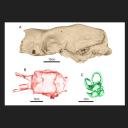
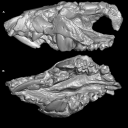
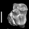
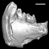
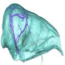
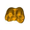
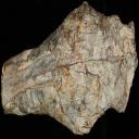
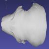
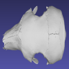
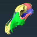
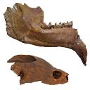
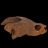
This contribution presents the three-dimensional digital models (i.e., skull, endocast, and inner ear) of a uniquely well-preserved and nearly complete skull (MCL 4228) attributed to the Late Pleistocene giant mylodontid ground sloth Ocnotherium giganteum, discovered in the Toca dos Ossos cave (Bahia State, Brazil). This specimen was described and figured in the following publication: Pujos et al. 2026: The neotropical giant ground sloth Ocnotherium giganteum (Xenarthra, Mylodontinae) from the Late Pleistocene of Brazil: anatomy, paleoneurology, and phylogenetic relationships. Zoological Journal of the Linnean Society. https://doi.org/10.1093/zoolinnean/zlag008
Ocnotherium giganteum MCL 4228 View specimen

|
M3#1870skull, endocast, inner ear Type: "3D_surfaces"doi: 10.18563/m3.sf.1870 state:in_press |
Download 3D surface file |
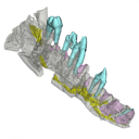
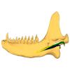
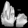
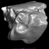
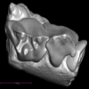
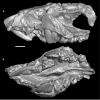
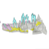
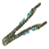
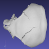
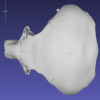
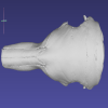
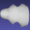
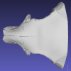
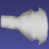
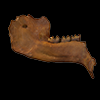
The present 3D Dataset contains the 3D models of the holotype and only specimen of Kalakocetus aurorae, a new cetacean retrieved from the Kalakot area in northwestern India. This specimen consists in a left hemimandible preserving the root of i3, p2, p4, m1 and m3 in situ. Its primitive morphology, with a tricuspid m3 morphologically intermediate between Raoellidae and Pakicetidae, makes it the first offshoot of Cetacea and provides crucial new elements to understand the setting up of the peculiar dental morphology of early cetaceans.
Kalakocetus aurorae GU/RJ/07 View specimen
|
M3#1803left hemi mandible with p2, p4, m1, m3 Type: "3D_surfaces"doi: 10.18563/m3.sf.1803 state:in_press |
Download 3D surface file |
|
M3#1804digitaly restored m1 Type: "3D_surfaces"doi: 10.18563/m3.sf.1804 state:in_press |
Download 3D surface file |
|
M3#1805digital restoration of complete mandible Type: "3D_surfaces"doi: 10.18563/m3.sf.1805 state:in_press |
Download 3D surface file |
|
M3#1810Scan Type: "3D_CT"doi: 10.18563/m3.sf.1810 state:in_press |
Download CT data |

The present 3D Dataset contains the 3D models of the two papionine remains found near Gabes and analyzed in Ksila et al. 2026 “A continental Messinian vertebrate fauna from the Ouedhref area, Southeast Tunisia.”
Macaca sp. UTM-O-Sa60 View specimen
|
M3#1872Right m1 or m2 Type: "3D_surfaces"doi: 10.18563/m3.sf.1872 state:in_press |
Download 3D surface file |
Macaca sp. UTM-O-Br6 View specimen
|
M3#1871Left upper canine Type: "3D_surfaces"doi: 10.18563/m3.sf.1871 state:in_press |
Download 3D surface file |
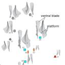
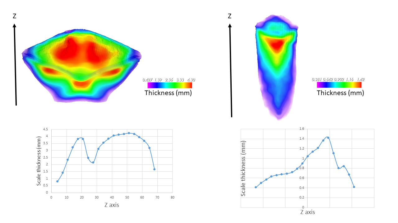
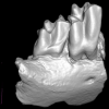
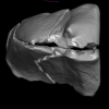
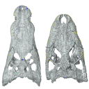
The present 3D dataset contains 15 specimens selected from the 69 3D models analyzed in the paper “3D topography as an indicator of change in food processing ability in the conodont genus Palmatolepis elements”. 3D topographic analysis of Palmatolepis P1 conodont elements from the Late Devonian period revealed an increase in blade sharpness together with a reduction in platform size. This indicates morphofunctional adaptation to more efficient prey processing.
Palmatolepis manticolepis UM CTB 144 View specimen

|
M3#1814Palmatolepis Manticolepis Type: "3D_surfaces"doi: 10.18563/m3.sf.1814 state:in_press |
Download 3D surface file |
Palmatolepis manticolepis UM CTB 151 View specimen

|
M3#1815Palmatolepis Manticolepis Type: "3D_surfaces"doi: 10.18563/m3.sf.1815 state:in_press |
Download 3D surface file |
Palmatolepis manticolepis UM CTB 078 View specimen

|
M3#1816Palmatolepis Manticolepis Type: "3D_surfaces"doi: 10.18563/m3.sf.1816 state:in_press |
Download 3D surface file |
Palmatolepis rhomboidea UM CTB 172 View specimen

|
M3#1817Palmatolepis rhomboidea Type: "3D_surfaces"doi: 10.18563/m3.sf.1817 state:in_press |
Download 3D surface file |
Palmatolepis glabra UM CTB 080 View specimen

|
M3#1818Palmatolepis glabra Type: "3D_surfaces"doi: 10.18563/m3.sf.1818 state:in_press |
Download 3D surface file |
Palmatolepis glabra UM CTB 177 View specimen

|
M3#1819Palmatolepis glabra Type: "3D_surfaces"doi: 10.18563/m3.sf.1819 state:in_press |
Download 3D surface file |
Palmatolepis glabra UM CTB 178 View specimen

|
M3#1820Palmatolepis glabra Type: "3D_surfaces"doi: 10.18563/m3.sf.1820 state:in_press |
Download 3D surface file |
Palmatolepis glabra UM CTB 179 View specimen

|
M3#1821Palmatolepis glabra Type: "3D_surfaces"doi: 10.18563/m3.sf.1821 state:in_press |
Download 3D surface file |
Palmatolepis gracilis UM CTB 186 View specimen

|
M3#1822Palmatolepis gracilis Type: "3D_surfaces"doi: 10.18563/m3.sf.1822 state:in_press |
Download 3D surface file |
Palmatolepis perlobata UM CTB 187 View specimen

|
M3#1823Palmatolepis perlobata Type: "3D_surfaces"doi: 10.18563/m3.sf.1823 state:in_press |
Download 3D surface file |
Palmatolepis perlobata UM CTB 189 View specimen

|
M3#1824Palmatolepis perlobata Type: "3D_surfaces"doi: 10.18563/m3.sf.1824 state:in_press |
Download 3D surface file |
Palmatolepis gracilis UM CTB 190 View specimen

|
M3#1825Palmatolepis gracilis Type: "3D_surfaces"doi: 10.18563/m3.sf.1825 state:in_press |
Download 3D surface file |
Palmatolepis perlobata UM CTB 191 View specimen

|
M3#1826Palmatolepis perlobata Type: "3D_surfaces"doi: 10.18563/m3.sf.1826 state:in_press |
Download 3D surface file |
Palmatolepis gracilis UM CTB 197 View specimen

|
M3#1827Palmatolepis gracilis Type: "3D_surfaces"doi: 10.18563/m3.sf.1827 state:in_press |
Download 3D surface file |
Palmatolepis gracilis UM CTB 200 View specimen

|
M3#1828Palmatolepis gracilis Type: "3D_surfaces"doi: 10.18563/m3.sf.1828 state:in_press |
Download 3D surface file |
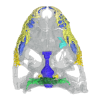
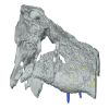
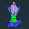
This contribution contains the 3D models of the paratympanic sinus system, the endocast and the neurovascular bony canal of the maxilla, premaxilla and the jugal of Leidyosuchus canadensis and Stangerochampsa mccabei described and figured in the following publication: G. Donzé, G. Perrichon, P. Vincent, JE. Martin, 2026. Comparative endocranial traits in the crocodylians Leidyosuchus canadensis and Stangerochampsa mccabei from the upper Cretaceous of Alberta, Canada. Journal of Anatomy.
Leidyosuchus canadensis TMP1986.221.1 View specimen

|
M3#1830Skull, endocast, paratympanic sinuses, jugal and maxillary neurovascular canals, alveoli Type: "3D_surfaces"doi: 10.18563/m3.sf.1830 state:in_press |
Download 3D surface file |
Stangerochampsa mccabei TMP1986.61.1 View specimen
|
M3#1831Skull, endocast, paratympanic sinuses, jugal and maxillary neurovascular canals, alveoli Type: "3D_surfaces"doi: 10.18563/m3.sf.1831 state:in_press |
Download 3D surface file |
Alligator mississipiensis OUVC 9761 View specimen

|
M3#1833Skull, endocast, paratympanic sinuses, jugal and maxillary neurovascular canals, alveolies Type: "3D_surfaces"doi: 10.18563/m3.sf.1833 state:in_press |
Download 3D surface file |
Alligator sinensis NHMW-Zoo-HS-37966 View specimen

|
M3#1835Skull, endocast, paratympanic sinus, jugal and maxillary neurovascular canals, alveoli Type: "3D_surfaces"doi: 10.18563/m3.sf.1835 state:in_press |
Download 3D surface file |
Crocodylus niloticus MHNL 50001399 View specimen

|
M3#1834Skull, endocast, paratympanic sinuses, jugal and maxillary neurovascular canals, alveolies Type: "3D_surfaces"doi: 10.18563/m3.sf.1834 state:in_press |
Download 3D surface file |
Diplocynodon ratelii LA86 View specimen
|
M3#1832Skull, endocast, paratympanic sinus, jugal and maxillary neurovascular canals, alveolies Type: "3D_surfaces"doi: 10.18563/m3.sf.1832 state:in_press |
Download 3D surface file |
This contribution contains the 3D models described and figured in: Phylogenetic signal in anteater snout morphology: implications for interpreting rare vermilinguan fossils. Palaeobiodiversity and Palaeoenvironments.
Indet indet VPPLT 977 View specimen

|
M3#17933D surface models of the cranium, nasal bone and cranial canals Type: "3D_surfaces"doi: 10.18563/m3.sf.1793 state:in_press |
Download 3D surface file |
Cyclopes didactylus M 1525 View specimen
|
M3#17943D models of the cranium and internal cranial canals Type: "3D_surfaces"doi: 10.18563/m3.sf.1794 state:in_press |
Download 3D surface file |
Cyclopes didactylus M 1571 View specimen
|
M3#17953D surface models of the cranium, nasal bone and cranial canals Type: "3D_surfaces"doi: 10.18563/m3.sf.1795 state:in_press |
Download 3D surface file |
Myrmecophaga tridactyla M 3023 View specimen
|
M3#17963D surface models of the cranium, nasal bone and cranial canals Type: "3D_surfaces"doi: 10.18563/m3.sf.1796 state:in_press |
Download 3D surface file |
Tamandua tetradactyla NHMUK 3.7.7.135 View specimen
|
M3#17973D models of the cranium and internal cranial canals Type: "3D_surfaces"doi: 10.18563/m3.sf.1797 state:in_press |
Download 3D surface file |
Tamandua tetradactyla NHMUK 4.7.4.90 View specimen
|
M3#17983D surface models of the cranium, nasal bone and cranial canals Type: "3D_surfaces"doi: 10.18563/m3.sf.1798 state:in_press |
Download 3D surface file |
Tamandua tetradactyla UM 788N View specimen
|
M3#17993D models of the cranium and internal cranial canals Type: "3D_surfaces"doi: 10.18563/m3.sf.1799 state:in_press |
Download 3D surface file |
Cyclopes didactylus MVZ 121210 View specimen
|
M3#18003D models of the cranium and internal cranial canals Type: "3D_surfaces"doi: 10.18563/m3.sf.1800 state:in_press |
Download 3D surface file |
Myrmecophaga tridactyla MVZ 112943 View specimen
|
M3#18013D models of the cranium and internal cranial canals Type: "3D_surfaces"doi: 10.18563/m3.sf.1801 state:in_press |
Download 3D surface file |
Myrmecophaga tridactyla MVZ 185238 View specimen
|
M3#18023D models of the cranium and internal cranial canals Type: "3D_surfaces"doi: 10.18563/m3.sf.1802 state:in_press |
Download 3D surface file |
|
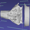
3D Printing an Explodable Dog Skull for Veterinary EducationWilliam C. Hooker
Published online: 17/12/2025 |

|
M3#1858PLYs of the segmented cranial bones with pre-fabricated magnetic casings and shelves for assembly following 3D printing Type: "3D_surfaces"doi: 10.18563/m3.sf.1858 state:published |
Download 3D surface file |
|
M3#1859PLYs of the segmented cranial bones of the "BOTTOM" cranial component. Downloadable for additional learning opportunities for students Type: "3D_surfaces"doi: 10.18563/m3.sf.1859 state:published |
Download 3D surface file |
|
3D models related to the publication: Révision des données sédimentologiques et biostratigraphiques des gisements à vertébrés des sables de l’Orléanais, à Beaugency, Tavers et Le Bardon (Miocène Moyen ; Loiret, France)Adrien de Perthuis
Published online: 31/10/2025 |
|
M3#1837Left upper M3 Type: "3D_surfaces"doi: 10.18563/m3.sf.1837 state:published |
Download 3D surface file |
Megamphicyon giganteus ULB-TAV-13 View specimen

|
M3#1531Left first lower molar Type: "3D_surfaces"doi: 10.18563/m3.sf.1531 state:published |
Download 3D surface file |
Hispanotherium matritense ULB-TAV-17 View specimen
|
M3#1532Left first lower molar Type: "3D_surfaces"doi: 10.18563/m3.sf.1532 state:published |
Download 3D surface file |
Plesiaceratherium lumiarense ULB-TAV-18 View specimen
|
M3#1533Left third upper molar Type: "3D_surfaces"doi: 10.18563/m3.sf.1533 state:published |
Download 3D surface file |
Chelydropsis aff. sansaniensis ULB-TAV-23 View specimen
|
M3#1535Cast of a skull Type: "3D_surfaces"doi: 10.18563/m3.sf.1535 state:published |
Download 3D surface file |
Ronzotherium romani ULB-TAV-4 View specimen
|
M3#1556Right fourth upper premolar Type: "3D_surfaces"doi: 10.18563/m3.sf.1556 state:published |
Download 3D surface file |
Prodeinotherium bavaricum ULB-TAV-24 View specimen
|
M3#1557left hemimandibule Type: "3D_surfaces"doi: 10.18563/m3.sf.1557 state:published |
Download 3D surface file |

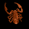
In this contribution a third new species of the rare genus Burmesescorpiops Lourenço, 2016 is described. The discovery of this new element belonging to the family Palaeoeuscorpiidae Lourenço, 2003 and to the subfamily Archaeoscorpiopinae Lourenço, 2015 brings further elements to support the validity of the genus Burmesescorpiops. This generic group remains however, poorly speciose. This is the latest discovery of Burmesescorpiops wunpawng, the name is derived from the Kachin Hilltribe peoples who are indigenous to the area. The data provided here is a 3D surface.
Burmesescorpiops wunpawng ps-gyi-01-25 View specimen
|
M3#18463d Surface Volume Type: "3D_surfaces"doi: 10.18563/m3.sf.1846 state:published |
Download 3D surface file |
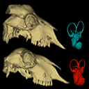
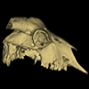
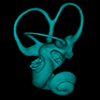
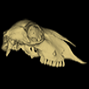
This contribution contains the 3D models described and figured in the following publication:Skull and Inner Ear Morphometrics in Sheep and Goats: Species and Breed Differentiation with Bioarchaeological Applications (Hemelsdael et al. submitted). The models include the external surface of a complete skull and inner ear of both a sheep (Ovis aries) and a goat (Capra hircus), generated from micro-CT scans. In the associated paper, we used 3D geometric morphometric data to assess inter and intra (i.e. between breeds) discrimination based on complete skulls, skull fragments and the semi-circular canals of the inner ear.
Capra hircus Amp_1 View specimen
|
M3#1806Skull of the goat Amp_1 Type: "3D_surfaces"doi: 10.18563/m3.sf.1806 state:published |
Download 3D surface file |
|
M3#1807Inner ear of the goat Amp_1 Type: "3D_surfaces"doi: 10.18563/m3.sf.1807 state:published |
Download 3D surface file |
Ovis aries UM_RR_2331 View specimen
|
M3#1808Skull of the sheep UM_RR_2331 Type: "3D_surfaces"doi: 10.18563/m3.sf.1808 state:published |
Download 3D surface file |
|
M3#1809Inner ear of the sheep UM_RR_2331 Type: "3D_surfaces"doi: 10.18563/m3.sf.1809 state:published |
Download 3D surface file |