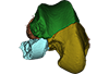
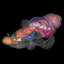
Explodable 3D Dog Skull for Veterinary Education
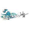
3D models of Ocnotherium skull
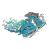
3D models of Kalakocetus, the earliest Cetacea
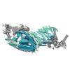
3D GM dataset of bird skeletal variation
Skeletal embryonic development in the catshark
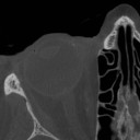
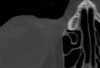
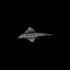
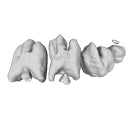
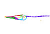
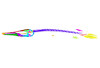
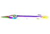
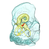
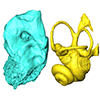
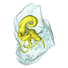
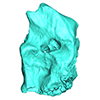
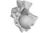
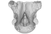
Bony connexions of the petrosal bone of extant hippos
bony labyrinth (14) , inner ear (11) , geometric morphometrics (10) , CT-scan (10) , Eocene (10) , Micro-CT (9) , Miocene (8)
Lionel Hautier (24) , Maëva Judith Orliac (23) , Laurent Marivaux (18) , Renaud Lebrun (14) , Rodolphe Tabuce (14) , Pierre-Olivier Antoine (13) , Bastien Mennecart (13)
|
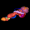
A human head micro-CT dataset : skull and soft tissuesGuillaume Captier
Published online: 10/05/2023 |
|
M3#1152Micro-ct data set Type: "3D_CT"doi: 10.18563/m3.sf.1152 state:published |
Download CT data |
We provide a 3D reconstruction of the skull of Latimeria chalumnae that can be easily accessed and visualized for a better understanding of its cranial anatomy. Different skeletal elements are saved as separate PLY files that can be combined to visualize the entire skull or isolated to virtually dissect the skull. We included some guidelines for a fast and easy visualization of the 3D skull.
Latimeria chalumnae MHNG 1080.070 View specimen

|
M3#1254the skeletal elements of the skull of Latimeria chalumnae included in 26 different PLY files Type: "3D_surfaces"doi: 10.18563/m3.sf.1254 state:published |
Download 3D surface file |
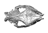
The present contribution contains the 3D virtual restoration of a Pliocene Lutrine right femur of Tobène, Senegal, described and figured in Lihoreau et al. (2021) : "A fossil terrestrial fauna from Tobène (Senegal) provides a unique early Pliocene window in Western Africa ". https://doi.org/10.1016/j.gr.2021.06.013
Indet indet SN-Tob-12-02 View specimen
|
M3#441Virtual restoration of SN-Tob-12-02 Type: "3D_surfaces"doi: 10.18563/m3.sf.441 state:published |
Download 3D surface file |
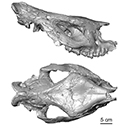
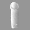
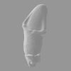
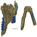
The present 3D Dataset contains the 3D model of the skull of the raoellid Indohyus indirae described in Patel et al. 2024.
Indohyus indirae RR 207 View specimen
|
M3#1259dorsoventrally crushed skull Type: "3D_surfaces"doi: 10.18563/m3.sf.1259 state:published |
Download 3D surface file |
Indohyus indirae RR 601 View specimen
|
M3#1268dorsoventrally crushed skull Type: "3D_surfaces"doi: 10.18563/m3.sf.1268 state:published |
Download 3D surface file |
This contribution provides for the first time the 3D model of the type specimen of Molassitherium delemontense (Mammalia, Rhinocerotidae) described in the following publication: Becker et al. (2013), Journal of Systematic Palaeontology, Vol. 11, Issue 8, 947–972, https://doi.org/10.1080/14772019.2012.699007. Conservation issues of the specimen and solutions using 3D model and 3D prints are detailed.
Molassitherium delemontense MJSN POI007–245 View specimen
|
M3#384Skull of Molassitherium delemontense Becker and Antoine, 2013 (in Becker et al. 2013): holotype Type: "3D_surfaces"doi: 10.18563/m3.sf.384 state:published |
Download 3D surface file |
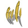
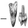
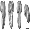
The present 3D dataset contains 3D models of new material from the middle Eocene of the Upper Subathu Formation in the Kalakot area (India), documenting the anterior dentition of the raoellid Indohyus indirae. Raoellidae are closely related to stem cetaceans and bring crucial information to understand the earliest phase of land to water transition in Cetacea.
Indohyus indirae GU/RJ/31 View specimen
|
M3#1505Right i1 Type: "3D_surfaces"doi: 10.18563/m3.sf.1505 state:published |
Download 3D surface file |
Indohyus indirae GU/RJ/32 View specimen
|
M3#1506Right i1 Type: "3D_surfaces"doi: 10.18563/m3.sf.1506 state:published |
Download 3D surface file |
Indohyus indirae GU/RJ/16 View specimen
|
M3#1507Left I3 Type: "3D_surfaces"doi: 10.18563/m3.sf.1507 state:published |
Download 3D surface file |
Indohyus indirae GU/RJ/23 View specimen
|
M3#1508Left I2 Type: "3D_surfaces"doi: 10.18563/m3.sf.1508 state:published |
Download 3D surface file |
Indohyus indirae GU/RJ/25 View specimen
|
M3#1509Left I1 Type: "3D_surfaces"doi: 10.18563/m3.sf.1509 state:published |
Download 3D surface file |
Indohyus indirae GU/RJ/26 View specimen
|
M3#1510Right I3 Type: "3D_surfaces"doi: 10.18563/m3.sf.1510 state:published |
Download 3D surface file |
Indohyus indirae GU/RJ/57 View specimen
|
M3#1511Left I1 Type: "3D_surfaces"doi: 10.18563/m3.sf.1511 state:published |
Download 3D surface file |
Indohyus indirae GU/RJ/61 View specimen
|
M3#1512Right upper canine Type: "3D_surfaces"doi: 10.18563/m3.sf.1512 state:published |
Download 3D surface file |
Indohyus indirae GU/RJ/63 View specimen
|
M3#1513Left upper canine Type: "3D_surfaces"doi: 10.18563/m3.sf.1513 state:published |
Download 3D surface file |
Indohyus indirae GU/RJ/74 View specimen
|
M3#1514Left upper canine Type: "3D_surfaces"doi: 10.18563/m3.sf.1514 state:published |
Download 3D surface file |
Indohyus indirae GU/RJ/439 View specimen
|
M3#1515Left upper canine Type: "3D_surfaces"doi: 10.18563/m3.sf.1515 state:published |
Download 3D surface file |
Indohyus indirae GU/RJ/457 View specimen
|
M3#1516Left upper canine Type: "3D_surfaces"doi: 10.18563/m3.sf.1516 state:published |
Download 3D surface file |
Indohyus indirae GU/RJ/846 View specimen
|
M3#1517Left upper canine Type: "3D_surfaces"doi: 10.18563/m3.sf.1517 state:published |
Download 3D surface file |
Indohyus indirae GU/RJ/822 View specimen
|
M3#1518Right fragmentary maxillary with decidual canine and I3 Type: "3D_surfaces"doi: 10.18563/m3.sf.1518 state:published |
Download 3D surface file |
Indohyus indirae GU/RJ/824 View specimen
|
M3#1519Right fragmentary mandible with lower canine and small part of the i3 Type: "3D_surfaces"doi: 10.18563/m3.sf.1519 state:published |
Download 3D surface file |
Indohyus indirae GU/RJ/838 View specimen
|
M3#1520Right fragmentary mandible with permanent i2, i3 and canine and small part of the root of the decidual i3 Type: "3D_surfaces"doi: 10.18563/m3.sf.1520 state:published |
Download 3D surface file |
Indohyus indirae GU/RJ/842 View specimen
|
M3#1521Left fragmentary mandible with decidual and permanent canine Type: "3D_surfaces"doi: 10.18563/m3.sf.1521 state:published |
Download 3D surface file |
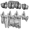
Indohyus indirae GU/RJ/26,32,56,57,457,822,838,842 : Composite Anterior dentition View specimen
|
M3#15293D composite reconstruction of the anterior dentition of Indohyus indirae with GU/RJ/57 (I1), 56 (I2), 26 (I3), 457 (Upper canine), 822 (P1), 32 (i1), 838 (i2, i3 and lower canine) and 842 (p1) Type: "3D_surfaces"doi: 10.18563/m3.sf.1529 state:published |
Download 3D surface file |
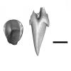
This contribution contains the three-dimensional models of the most complete and/or informative fossil materials attributed to Peradectes crocheti Gernelle, 2024, the earliest peradectid metatherian species of Europe, from its type locality (Palette, Provence, ~55 Ma). These specimens were analyzed and discussed in: Gernelle et al. (2024), Taxonomy and evolutionary history of peradectids (Metatheria): new data from the early Eocene of France. https://doi.org/10.1007/s10914-024-09724-5
Peradectes crocheti MHN.AIX.PV.2018.26.14 View specimen
|
M3#14993D surface model of MHN.AIX.PV.2018.26.14, fragmentary left maxilla with C-P1, anterior root of P2, and M1-M3 Type: "3D_surfaces"doi: 10.18563/m3.sf.1499 state:published |
Download 3D surface file |
Peradectes crocheti MHN.AIX.PV.2017.6.6 View specimen
|
M3#15003D surface model of MHN.AIX.PV.2017.6.6, left P2 Type: "3D_surfaces"doi: 10.18563/m3.sf.1500 state:published |
Download 3D surface file |
Peradectes crocheti MHN.AIX.PV.2017.6.7 View specimen
|
M3#15013D surface model of MHN.AIX.PV.2017.6.7, left M3 Type: "3D_surfaces"doi: 10.18563/m3.sf.1501 state:published |
Download 3D surface file |
Peradectes crocheti MHN.AIX.PV.2017.6.8 View specimen
|
M3#15023D surface model of MHN.AIX.PV.2017.6.8, right hemi-mandible fragment with canine alveolus, posterior root of p1, partial p2, p3, partial m1, and m2-m3 Type: "3D_surfaces"doi: 10.18563/m3.sf.1502 state:published |
Download 3D surface file |
Peradectes crocheti MHN.AIX.PV.2017.6.9 View specimen
|
M3#15033D surface model of MHN.AIX.PV.2017.6.9, leftm1-m4 row with fragments of dentary Type: "3D_surfaces"doi: 10.18563/m3.sf.1503 state:published |
Download 3D surface file |
Peradectes crocheti MHN.AIX.PV.2017.6.14 View specimen
|
M3#15043D surface model of MHN.AIX.PV.2017.6.14, right astragalus Type: "3D_surfaces"doi: 10.18563/m3.sf.1504 state:published |
Download 3D surface file |
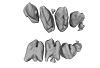
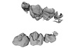
The present 3D Dataset contains the 3D models analyzed in Assemat et al. 2023: Shape diversity in conodont elements, a quantitative study using 3D topography. Marine Micropaleontology 184. https://doi.org/10.1016/j.marmicro.2023.102292
P1 elements represent dental components of the conodont apparatus that perform the final stage of food processing before ingestion. Consequently, quantifying the shape of P1 elements across the topographic indices of different conodont species becomes crucial for deciphering the diversity in feeding behavior within this group.
Bispathodus aculeatus UM CTB 082 View specimen
|
M3#1404P element Type: "3D_surfaces"doi: 10.18563/m3.sf.1404 state:published |
Download 3D surface file |
Bispathodus aculeatus UM CTB 083 View specimen

|
M3#1405P element Type: "3D_surfaces"doi: 10.18563/m3.sf.1405 state:published |
Download 3D surface file |
Bispathodus aculeatus UM CTB 086 View specimen
|
M3#1406P element Type: "3D_surfaces"doi: 10.18563/m3.sf.1406 state:published |
Download 3D surface file |
Bispathodus ultimus UM CTB 088 View specimen

|
M3#1407P element Type: "3D_surfaces"doi: 10.18563/m3.sf.1407 state:published |
Download 3D surface file |
Bispathodus aculeatus UM CTB 089 View specimen
|
M3#1408P element Type: "3D_surfaces"doi: 10.18563/m3.sf.1408 state:published |
Download 3D surface file |
Bispathodus costatus UM CTB 090 View specimen
|
M3#1409P element Type: "3D_surfaces"doi: 10.18563/m3.sf.1409 state:published |
Download 3D surface file |
Bispathodus ultimus UM CTB 092 View specimen
|
M3#1410P element Type: "3D_surfaces"doi: 10.18563/m3.sf.1410 state:published |
Download 3D surface file |
Bispathodus costatus UM CTB 093 View specimen
|
M3#1411P element Type: "3D_surfaces"doi: 10.18563/m3.sf.1411 state:published |
Download 3D surface file |
Bispathodus spinulicostatus UM CTB 094 View specimen
|
M3#1412P element Type: "3D_surfaces"doi: 10.18563/m3.sf.1412 state:published |
Download 3D surface file |
Bispathodus aculeatus UM CTB 096 View specimen

|
M3#1413P element Type: "3D_surfaces"doi: 10.18563/m3.sf.1413 state:published |
Download 3D surface file |
Bispathodus ultimus UM CTB 098 View specimen
|
M3#1414P element Type: "3D_surfaces"doi: 10.18563/m3.sf.1414 state:published |
Download 3D surface file |
Bispathodus costatus UM CTB 060 View specimen
|
M3#1415P element Type: "3D_surfaces"doi: 10.18563/m3.sf.1415 state:published |
Download 3D surface file |
Bispathodus spinulicostatus UM CTB 073 View specimen

|
M3#1416P element Type: "3D_surfaces"doi: 10.18563/m3.sf.1416 state:published |
Download 3D surface file |
Branmehla suprema UM CTB 049 View specimen

|
M3#1417P element Type: "3D_surfaces"doi: 10.18563/m3.sf.1417 state:published |
Download 3D surface file |
Branmehla inornata UM CTB 100 View specimen
|
M3#1418P element Type: "3D_surfaces"doi: 10.18563/m3.sf.1418 state:published |
Download 3D surface file |
Bispathodus stabilis (morphe 1) UM CTB 101 View specimen
|
M3#1419P element Type: "3D_surfaces"doi: 10.18563/m3.sf.1419 state:published |
Download 3D surface file |
Branmehla suprema UM CTB 102 View specimen

|
M3#1420P element Type: "3D_surfaces"doi: 10.18563/m3.sf.1420 state:published |
Download 3D surface file |
Branmehla suprema UM CTB 103 View specimen

|
M3#1421P element Type: "3D_surfaces"doi: 10.18563/m3.sf.1421 state:published |
Download 3D surface file |
Branmehla suprema UM CTB 104 View specimen

|
M3#1422P element Type: "3D_surfaces"doi: 10.18563/m3.sf.1422 state:published |
Download 3D surface file |
Branmehla suprema UM CTB 105 View specimen
|
M3#1423P element Type: "3D_surfaces"doi: 10.18563/m3.sf.1423 state:published |
Download 3D surface file |
Branmehla suprema UM CTB 106 View specimen
|
M3#1424P element Type: "3D_surfaces"doi: 10.18563/m3.sf.1424 state:published |
Download 3D surface file |
Branmehla suprema UM CTB 072 View specimen

|
M3#1425P element Type: "3D_surfaces"doi: 10.18563/m3.sf.1425 state:published |
Download 3D surface file |
Branmehla suprema UM CTB 107 View specimen
|
M3#1426P element Type: "3D_surfaces"doi: 10.18563/m3.sf.1426 state:published |
Download 3D surface file |
Branmehla suprema UM CTB 108 View specimen

|
M3#1427P element Type: "3D_surfaces"doi: 10.18563/m3.sf.1427 state:published |
Download 3D surface file |
Branmehla suprema UM CTB 109 View specimen
|
M3#1428P element Type: "3D_surfaces"doi: 10.18563/m3.sf.1428 state:published |
Download 3D surface file |
Bispathodus stabilis (morphe 1) UM CTB 110 View specimen

|
M3#1429P element Type: "3D_surfaces"doi: 10.18563/m3.sf.1429 state:published |
Download 3D surface file |
Palmatolepis gracilis UM CTB 112 View specimen
|
M3#1430P element Type: "3D_surfaces"doi: 10.18563/m3.sf.1430 state:published |
Download 3D surface file |
Palmatolepis gracilis UM CTB 061 View specimen

|
M3#1431P element Type: "3D_surfaces"doi: 10.18563/m3.sf.1431 state:published |
Download 3D surface file |
Palmatolepis gracilis UM CTB 115 View specimen
|
M3#1432P element Type: "3D_surfaces"doi: 10.18563/m3.sf.1432 state:published |
Download 3D surface file |
Palmatolepis gracilis UM CTB 116 View specimen
|
M3#1433P element Type: "3D_surfaces"doi: 10.18563/m3.sf.1433 state:published |
Download 3D surface file |
Palmatolepis gracilis UM CTB 117 View specimen
|
M3#1434P element Type: "3D_surfaces"doi: 10.18563/m3.sf.1434 state:published |
Download 3D surface file |
Palmatolepis gracilis UM CTB 062 View specimen

|
M3#1435P element Type: "3D_surfaces"doi: 10.18563/m3.sf.1435 state:published |
Download 3D surface file |
Palmatolepis gracilis UM CTB 118 View specimen

|
M3#1436P element Type: "3D_surfaces"doi: 10.18563/m3.sf.1436 state:published |
Download 3D surface file |
Palmatolepis gracilis UM CTB 119 View specimen
|
M3#1437P element Type: "3D_surfaces"doi: 10.18563/m3.sf.1437 state:published |
Download 3D surface file |
Palmatolepis gracilis UM CTB 120 View specimen
|
M3#1438P element Type: "3D_surfaces"doi: 10.18563/m3.sf.1438 state:published |
Download 3D surface file |
Polygnathus communis UM CTB 075 View specimen
|
M3#1439P element Type: "3D_surfaces"doi: 10.18563/m3.sf.1439 state:published |
Download 3D surface file |
Polygnathus communis UM CTB 121 View specimen
|
M3#1440P element Type: "3D_surfaces"doi: 10.18563/m3.sf.1440 state:published |
Download 3D surface file |
Polygnathus communis UM CTB 122 View specimen

|
M3#1441P element Type: "3D_surfaces"doi: 10.18563/m3.sf.1441 state:published |
Download 3D surface file |
Polygnathus communis UM CTB 123 View specimen
|
M3#1442P element Type: "3D_surfaces"doi: 10.18563/m3.sf.1442 state:published |
Download 3D surface file |
Polygnathus communis UM CTB 125 View specimen
|
M3#1443P element Type: "3D_surfaces"doi: 10.18563/m3.sf.1443 state:published |
Download 3D surface file |
Polygnathus communis UM CTB 126 View specimen
|
M3#1444P element Type: "3D_surfaces"doi: 10.18563/m3.sf.1444 state:published |
Download 3D surface file |
Polygnathus communis UM CTB 128 View specimen
|
M3#1445P element Type: "3D_surfaces"doi: 10.18563/m3.sf.1445 state:published |
Download 3D surface file |
Polygnathus communis UM CTB 130 View specimen
|
M3#1446P element Type: "3D_surfaces"doi: 10.18563/m3.sf.1446 state:published |
Download 3D surface file |
Polygnathus communis UM CTB 131 View specimen
|
M3#1447P element Type: "3D_surfaces"doi: 10.18563/m3.sf.1447 state:published |
Download 3D surface file |
Polygnathus communis UM CTB 132 View specimen
|
M3#1448P element Type: "3D_surfaces"doi: 10.18563/m3.sf.1448 state:published |
Download 3D surface file |
Polygnathus communis UM CTB 133 View specimen
|
M3#1449P element Type: "3D_surfaces"doi: 10.18563/m3.sf.1449 state:published |
Download 3D surface file |
Polygnathus symmetricus UM CTB 139 View specimen
|
M3#1450P element Type: "3D_surfaces"doi: 10.18563/m3.sf.1450 state:published |
Download 3D surface file |
Polygnathus symmetricus UM CTB 140 View specimen
|
M3#1451P element Type: "3D_surfaces"doi: 10.18563/m3.sf.1451 state:published |
Download 3D surface file |
Polygnathus symmetricus UM CTB 141 View specimen
|
M3#1452P element Type: "3D_surfaces"doi: 10.18563/m3.sf.1452 state:published |
Download 3D surface file |
Polygnathus symmetricus UM CTB 142 View specimen
|
M3#1453P element Type: "3D_surfaces"doi: 10.18563/m3.sf.1453 state:published |
Download 3D surface file |
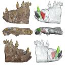
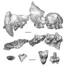
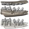
This contribution contains the 3D models described and figured in the following publication: Gaetano, L. C., Abdala, F., Mancuso, C, and Vega N.2025. New traversodontid cynodont from the Late Triassic Chañares Formation. Publicación Electrónica de la Asociación Paleontológica Argentina.
Pontognathus ignotus PULR-V 287 View specimen
|
M3#1647partial snout preserving the lateralmost incisor, the base of the canine, and several postcanines Type: "3D_surfaces"doi: 10.18563/m3.sf.1647 state:published |
Download 3D surface file |
Massetognathus pascuali PULR-V 289 View specimen
|
M3#1646partial lower jaw Type: "3D_surfaces"doi: 10.18563/m3.sf.1646 state:published |
Download 3D surface file |
The present 3D Dataset contains the 3D models analyzed in: Hirose, A., Nakashima, T., Yamada, S., Uwabe, C., Kose, K., Takakuwa, T. 2012. Embryonic liver morphology and morphometry by magnetic resonance microscopic imaging. Anat Rec (Hoboken) 295, 51-59. doi: 10.1002/ar.21496
Homo sapiens KC-CS14LIV1387 View specimen
|
M3#64Human liver at Carnegie Stage (CS) 14 Type: "3D_surfaces"doi: 10.18563/m3.sf.64 state:published |
Download 3D surface file |
Homo sapiens KC-CS15LIV5074 View specimen
|
M3#65Human liver at Carnegie Stage (CS) 15 Type: "3D_surfaces"doi: 10.18563/m3.sf.65 state:published |
Download 3D surface file |
Homo sapiens KC-CS16LIV2578 View specimen
|
M3#66Human liver at Carnegie Stage (CS) 16 Type: "3D_surfaces"doi: 10.18563/m3.sf.66 state:published |
Download 3D surface file |
Homo sapiens KC-CS17LIV17832 View specimen
|
M3#67Human liver at Carnegie Stage (CS) 17 Type: "3D_surfaces"doi: 10.18563/m3.sf.67 state:published |
Download 3D surface file |
Homo sapiens KC-CS18LIV21124 View specimen
|
M3#68Human liver at Carnegie Stage (CS) 18 Type: "3D_surfaces"doi: 10.18563/m3.sf.68 state:published |
Download 3D surface file |
Homo sapiens KC-CS19LIV14353 View specimen

|
M3#69Human liver at Carnegie Stage (CS) 19 Type: "3D_surfaces"doi: 10.18563/m3.sf.69 state:published |
Download 3D surface file |
Homo sapiens KC-CS20LIV20701 View specimen

|
M3#70Human liver at Carnegie Stage (CS) 20 Type: "3D_surfaces"doi: 10.18563/m3.sf.70 state:published |
Download 3D surface file |
Homo sapiens KC-CS21LIV25858 View specimen
|
M3#71Human liver at Carnegie Stage (CS) 21 Type: "3D_surfaces"doi: 10.18563/m3.sf.71 state:published |
Download 3D surface file |
Homo sapiens KC-CS22LIV22226 View specimen
|
M3#72Human liver at Carnegie Stage (CS) 22 Type: "3D_surfaces"doi: 10.18563/m3.sf.72 state:published |
Download 3D surface file |
Homo sapiens KC-CS23LIV25704 View specimen
|
M3#73Human liver at Carnegie Stage (CS) 23 Type: "3D_surfaces"doi: 10.18563/m3.sf.73 state:published |
Download 3D surface file |

This contribution contains 3D models of the cranial endoskeleton of three specimens of the Permian ‘acanthodian’ stem-group chondrichthyan (cartilaginous fish) Acanthodes confusus, obtained using computed tomography. These datasets were described and analyzed in Dearden et al. (2024) “3D models related to the publication: The pharynx of the iconic stem-group chondrichthyan Acanthodes Agassiz, 1833 revisited with micro computed tomography.” Zoological Journal of the Linnean Society
Acanthodes confusus MNHN-F-SAA20 View specimen
|
M3#14703D surfaces representing the three-dimensionally fossilised head of Acanthodes confusus Type: "3D_surfaces"doi: 10.18563/m3.sf.1470 state:published |
Download 3D surface file |
Acanthodes confusus MNHN-F-SAA21 View specimen
|
M3#14713D surfaces representing the three-dimensionally fossilised head of Acanthodes confusus Type: "3D_surfaces"doi: 10.18563/m3.sf.1471 state:published |
Download 3D surface file |
Acanthodes confusus MNHN-F-SAA24 View specimen
|
M3#14723D surfaces representing the three-dimensionally fossilised head of Acanthodes confusus Type: "3D_surfaces"doi: 10.18563/m3.sf.1472 state:published |
Download 3D surface file |
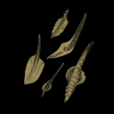
The present 3D Dataset contains the 3D models analyzed in ”Morphological features of tooth development and replacement in the rabbit Oryctolagus cuniculus”, Archives of Oral Biology, https://doi.org/10.1016/j.archoralbio.2019.104576
Oryctogalus cuniculus E14 View specimen
|
M3#390Right cheek teeth, Left and right incisors at 14 dpf Type: "3D_surfaces"doi: 10.18563/m3.sf.390 state:published |
Download 3D surface file |
Oryctogalus cuniculus E16 View specimen
|
M3#391Left cheek teeth, Left and right incisors at 16 dpf Type: "3D_surfaces"doi: 10.18563/m3.sf.391 state:published |
Download 3D surface file |
Oryctogalus cuniculus E18 View specimen
|
M3#392Left cheek teeth and incisors at 18 dpf Type: "3D_surfaces"doi: 10.18563/m3.sf.392 state:published |
Download 3D surface file |
Oryctogalus cuniculus E20 View specimen
|
M3#393Left cheek teeth and incisors at 20 dpf Type: "3D_surfaces"doi: 10.18563/m3.sf.393 state:published |
Download 3D surface file |
Oryctogalus cuniculus E22 View specimen
|
M3#394Left lower cheek teeth and incisors, right upper cheek teeth and incisors at 22 dpf Type: "3D_surfaces"doi: 10.18563/m3.sf.394 state:published |
Download 3D surface file |
Oryctogalus cuniculus E24 View specimen
|
M3#395Left cheek teeth and incisors at 24 dpf Type: "3D_surfaces"doi: 10.18563/m3.sf.395 state:published |
Download 3D surface file |
Oryctogalus cuniculus E28 View specimen
|
M3#396Right cheek teeth and incisors at 28 dpf Type: "3D_surfaces"doi: 10.18563/m3.sf.396 state:published |
Download 3D surface file |
Oryctogalus cuniculus E26 View specimen
|
M3#397Right cheek teeth and incisors at 26 dpf Type: "3D_surfaces"doi: 10.18563/m3.sf.397 state:published |
Download 3D surface file |
Using X-ray microtomography, we describe the ossification events during the larval development of a non-teleost actinopterygian species: the Cuban gar Atractosteus tristoechus from the order Lepisosteiformes. We provide a detailed developmental series for each anatomical structure, covering a large sequence of mineralization events going from an early stage (13 days post-hatching, 21mm total length) to an almost fully ossified larval stage (118dph or 87mm in standard length). With this work, we expect to bring new developmental data to be used in further comparative studies with other lineages of bony vertebrates. We also hope that the on-line publication of these twelve successive 3D reconstructions, fully labelled and flagged, will be an educational tool for all students in comparative anatomy.
Atractosteus tristoechus At1-13dph View specimen

|
M3#94At1-13dph : 13 dph larvae, 21 mm TL Type: "3D_surfaces"doi: 10.18563/m3.sf.94 state:published |
Download 3D surface file |
Atractosteus tristoechus At2-16dph View specimen
|
M3#95Atractosteus tristoechus larva, 16 dph, 26mm SL. Type: "3D_surfaces"doi: 10.18563/m3.sf.95 state:published |
Download 3D surface file |
Atractosteus tristoechus At3-19dph View specimen

|
M3#96Atractosteus tristoechus larva, 19 dph, 27mm SL. Type: "3D_surfaces"doi: 10.18563/m3.sf.96 state:published |
Download 3D surface file |
Atractosteus tristoechus At4-22dph View specimen
|
M3#97Atractosteus tristoechus larva, 22dph, 30mm SL. Type: "3D_surfaces"doi: 10.18563/m3.sf.97 state:published |
Download 3D surface file |
Atractosteus tristoechus At5-26dph View specimen
|
M3#98Atractosteus tristoechus larva, 26 dph, 32mm SL. Type: "3D_surfaces"doi: 10.18563/m3.sf.98 state:published |
Download 3D surface file |
Atractosteus tristoechus At6-31dph View specimen
|
M3#99Atractosteus tristoechus larva, 31 dph, 39mm SL. Type: "3D_surfaces"doi: 10.18563/m3.sf.99 state:published |
Download 3D surface file |
Atractosteus tristoechus At7-37dph View specimen
|
M3#100Atractosteus tristoechus larva, 37 dph, 43mm SL. Type: "3D_surfaces"doi: 10.18563/m3.sf.100 state:published |
Download 3D surface file |
Atractosteus tristoechus At8-52dph View specimen
|
M3#101Atractosteus tristoechus larva, 52 dph, 46mm SL. Type: "3D_surfaces"doi: 10.18563/m3.sf.101 state:published |
Download 3D surface file |
Atractosteus tristoechus At9-74dph View specimen
|
M3#102Atractosteus tristoechus larva, 74 dph, 61mm SL. Not all structures are colored, only newly ossified ones. Type: "3D_surfaces"doi: 10.18563/m3.sf.102 state:published |
Download 3D surface file |
Atractosteus tristoechus At10-89dph View specimen
|
M3#103Atractosteus tristoechus larva, 89 dph, 63mm SL. Not all structures are colored, only newly ossified ones. You may find the tag file in the At1-13dph reconstruction data. Type: "3D_surfaces"doi: 10.18563/m3.sf.103 state:published |
Download 3D surface file |
Atractosteus tristoechus At11-104dph View specimen
|
M3#104Atractosteus tristoechus larva, 104 dph, 70mm SL. Not all structures are colored, only newly ossified ones. Type: "3D_surfaces"doi: 10.18563/m3.sf.104 state:published |
Download 3D surface file |
Atractosteus tristoechus At12-118dph View specimen
|
M3#105Atractosteus tristoechus larva, 118 dph, 87mm SL. Type: "3D_surfaces"doi: 10.18563/m3.sf.105 state:published |
Download 3D surface file |
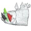
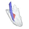
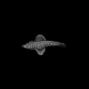
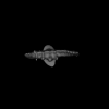
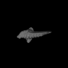
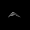
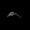
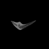
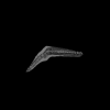
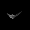
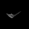
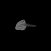
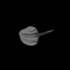
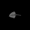
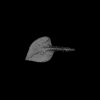
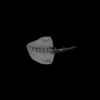
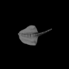
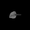
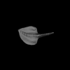
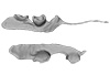
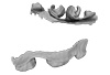
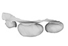
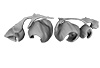
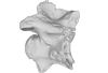
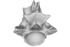
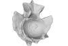
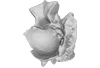
The present 3D Dataset contains the 3D models analyzed in Mennecart, B., Duranthon, F., & Costeur, L. 2024. Systematic contribution of the auditory region to the knowledge of the oldest European Bovidae (Mammalia, Ruminantia). Journal of Anatomy XXX. https://doi.org/10.1111/joa.14132
Pusillutragus montrealensis MHNT.PAL.2015.0.2261.4 View specimen
|
M3#1522Right petrosal, bony labyrinth, stapes Type: "3D_surfaces"doi: 10.18563/m3.sf.1522 state:published |
Download 3D surface file |
Pusillutragus montrealensis MHNT.PAL.2015.0.2261.9 View specimen

|
M3#1523Left petrosal and left bony labyrinth Type: "3D_surfaces"doi: 10.18563/m3.sf.1523 state:published |
Download 3D surface file |
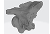
Eotragus artenensis SMNS-P-41625 View specimen
|
M3#1524Petrosal (right), bony labyrinth (left) Type: "3D_surfaces"doi: 10.18563/m3.sf.1524 state:published |
Download 3D surface file |
Eotragus clavatus NMB San.15056 View specimen
|
M3#1528Right petrosal and right bony labyrinth Type: "3D_surfaces"doi: 10.18563/m3.sf.1528 state:published |
Download 3D surface file |
Eotragus clavatus NMB San.15055 View specimen
|
M3#1526Left Petrosal Type: "3D_surfaces"doi: 10.18563/m3.sf.1526 state:published |
Download 3D surface file |
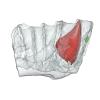
The present 3D dataset contains the 3D models analyzed in the publication: Rosa, R. M., Salvador, R. B., & Cavallari, D. C. (2025). The disappearing act of the magician tree snail: anatomy, distribution, and phylogenetic relationships of Drymaeus magus (Gastropoda: Bulimulidae), a long-lost species hidden in plain sight. Zoological Journal of the Linnean Society.
Drymaeus magus CMRP 1049 View specimen
|
M3#1597Internal organs of Drymaeus magus Type: "3D_surfaces"doi: 10.18563/m3.sf.1597 state:published |
Download 3D surface file |

|
M3#1598External surface of Drymaeus magus Type: "3D_surfaces"doi: 10.18563/m3.sf.1598 state:published |
Download 3D surface file |
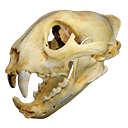
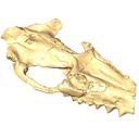
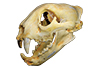
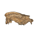
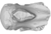
The present 3D Dataset contains the 3D model of a skull analyzed in “A Puma concolor (Carnivora: Felidae) in the Middle-Late Holocene landscapes of the Brazilian Northeast (Bahia): submerged cave deposits and stable isotopes”. The 3D model was generated by photogrammetry.
Puma concolor MN 57461 View specimen
|
M3#843Cranium Type: "3D_surfaces"doi: 10.18563/m3.sf.843 state:published |
Download 3D surface file |
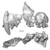
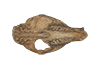
The present 3D Dataset contains the 3D model of a specimen of Metamynodon planifrons (UNISTRA.2015.0.1106) described and figured in: Veine-Tonizzo, L., Tissier, J., Bukhsianidze, M., Vasilyan, D., Becker, D., 2023, Cranial morphology and phylogenetic relationships of Amynodontidae Scott & Osborn, 1883 (Perissodactyla, Rhinocerotoidea).
Metamynodon planifrons UNISTRA.2015.0.1106 View specimen
|
M3#716Textured 3D surface model of the skull of the specimen UNISTRA.2015.0.1106 with right C1 and both rows of P2-M3. Type: "3D_surfaces"doi: 10.18563/m3.sf.716 state:published |
Download 3D surface file |
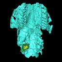
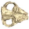
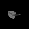
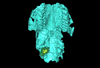
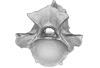
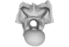
The present 3D Dataset contains the 3D models of the holotype (NMB Sth. 833) of the new species Micromeryx? eiselei analysed in the article Aiglstorfer, M., Costeur, L., Mennecart, B., Heizmann, E.P.J.. 2017. Micromeryx? eiselei - a new moschid species from Steinheim am Albuch, Germany, and the first comprehensive description of moschid cranial material from the Miocene of Central Europe. PlosOne https://doi.org/10.1371/journal.pone.0185679
Micromeryx? eiselei NMB Sth. 833 View specimen
|
M3#284The 3 D surfaces comprises the skull, petrosal, and bony labyrinth of NMB Sth.833, the holotype of Micromeryx? eiselei Type: "3D_surfaces"doi: 10.18563/m3.sf.284 state:published |
Download 3D surface file |

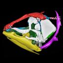
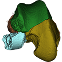
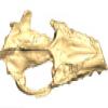
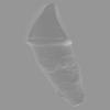
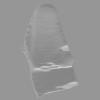
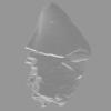
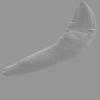
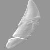
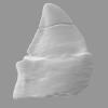
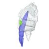
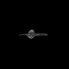
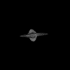
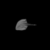
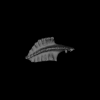
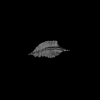
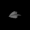
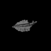
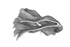
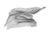
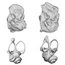
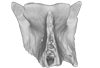
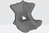
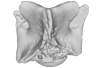
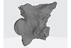
This project presents the 3D models of two isolated petrosals from the Oligocene locality of Pech de Fraysse (Quercy, France) here attributed to the genus Prodremotherium Filhol, 1877. Our aim is to describe the petrosal morphology of this Oligocene “early ruminant” as only few data are available in the literature for Oligocene taxa.
Prodremotherium sp. UM PFY 4053 View specimen

|
M3#7Labelled 3D model of right isolated petrosal of Prodremotherium sp. from Pech de Fraysse (Quercy, MP 28) Type: "3D_surfaces"doi: 10.18563/m3.sf7 state:published |
Download 3D surface file |
Prodremotherium sp. UM PFY 4054 View specimen

|
M3#8Labelled 3D model of right isolated petrosal of Prodremotherium sp. from Pech de Fraysse (Quercy, MP 28) Type: "3D_surfaces"doi: 10.18563/m3.sf8 state:published |
Download 3D surface file |
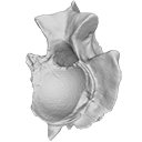
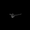
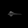
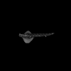
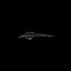
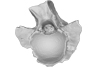
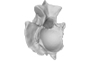
This contribution contains the 3D models described and figured in the following publication: Georgalis, G.L., G. Guinot, K.E. Kassegne, Y.Z. Amoudji, A.K.C. Johnson, H. Cappetta and L. Hautier. 2021. An assemblage of giant aquatic snakes (Serpentes, Palaeophiidae) from the Eocene of Togo. Swiss Journal of Palaeontology 140, https://doi.org/10.1186/s13358-021-00236-w
Palaeophis africanus UM KPO 21 View specimen
|
M3#821Trunk vertebra UM KPO 21 of Palaeophis africanus Type: "3D_surfaces"doi: 10.18563/m3.sf.821 state:published |
Download 3D surface file |
Palaeophis africanus UM KPO 22 View specimen
|
M3#822Trunk vertebra UM KPO 22 of Palaeophis africanus from the Eocene of Togo Type: "3D_surfaces"doi: 10.18563/m3.sf.822 state:published |
Download 3D surface file |
Palaeophis africanus UM KPO 23 View specimen
|
M3#823Trunk vertebra UM KPO 23 of Palaeophis africanus Type: "3D_surfaces"doi: 10.18563/m3.sf.823 state:published |
Download 3D surface file |
Palaeophis africanus UM KPO 24 View specimen
|
M3#824Trunk vertebra UM KPO 24 of Palaeophis africanus Type: "3D_surfaces"doi: 10.18563/m3.sf.824 state:published |
Download 3D surface file |
Palaeophis africanus UM KPO 25 View specimen
|
M3#825Trunk vertebra UM KPO 25 of Palaeophis africanus Type: "3D_surfaces"doi: 10.18563/m3.sf.825 state:published |
Download 3D surface file |
Palaeophis africanus UM KPO 26 View specimen
|
M3#826Trunk vertebra UM KPO 26 of Palaeophis africanus Type: "3D_surfaces"doi: 10.18563/m3.sf.826 state:published |
Download 3D surface file |
Palaeophis africanus UM KPO 27 View specimen
|
M3#827Trunk vertebra UM KPO 27 of Palaeophis africanus Type: "3D_surfaces"doi: 10.18563/m3.sf.827 state:published |
Download 3D surface file |
Palaeophis africanus UM KPO 28 View specimen
|
M3#828Trunk vertebra UM KPO 28 of Palaeophis africanus Type: "3D_surfaces"doi: 10.18563/m3.sf.828 state:published |
Download 3D surface file |
Palaeophis africanus UM KPO 29 View specimen
|
M3#829Trunk vertebra UM KPO 29 of Palaeophis africanus Type: "3D_surfaces"doi: 10.18563/m3.sf.829 state:published |
Download 3D surface file |
Palaeophis africanus UM KPO 30 View specimen
|
M3#830Trunk vertebra UM KPO 30 of Palaeophis africanus Type: "3D_surfaces"doi: 10.18563/m3.sf.830 state:published |
Download 3D surface file |
Palaeophis africanus UM KPO 31 View specimen
|
M3#831Trunk vertebra UM KPO 28 of Palaeophis africanus Type: "3D_surfaces"doi: 10.18563/m3.sf.831 state:published |
Download 3D surface file |
Palaeophis africanus UM KPO 32 View specimen
|
M3#832Trunk vertebra UM KPO 32 of Palaeophis africanus Type: "3D_surfaces"doi: 10.18563/m3.sf.832 state:published |
Download 3D surface file |
Palaeophis africanus UM KPO 33 View specimen
|
M3#833Trunk vertebra UM KPO 33 of Palaeophis africanus Type: "3D_surfaces"doi: 10.18563/m3.sf.833 state:published |
Download 3D surface file |
Palaeophis africanus UM KPO 34 View specimen
|
M3#839Trunk vertebra UM KPO 34 of Palaeophis africanus Type: "3D_surfaces"doi: 10.18563/m3.sf.839 state:published |
Download 3D surface file |
Palaeophis africanus UM KPO 35 View specimen
|
M3#840Trunk vertebra UM KPO 35 of Palaeophis africanus Type: "3D_surfaces"doi: 10.18563/m3.sf.840 state:published |
Download 3D surface file |
Palaeophis africanus UM KPO 36 View specimen
|
M3#841Trunk vertebra UM KPO 36 of Palaeophis africanus Type: "3D_surfaces"doi: 10.18563/m3.sf.841 state:published |
Download 3D surface file |
Palaeophis africanus UM KPO 37 View specimen
|
M3#842Trunk vertebra UM KPO 37 of Palaeophis africanus Type: "3D_surfaces"doi: 10.18563/m3.sf.842 state:published |
Download 3D surface file |


