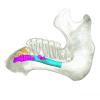
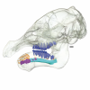
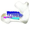
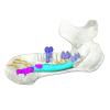
Explodable 3D Dog Skull for Veterinary Education
3D models of Ocnotherium skull
3D models of Kalakocetus, the earliest Cetacea
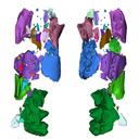
3D GM dataset of bird skeletal variation
Skeletal embryonic development in the catshark
Bony connexions of the petrosal bone of extant hippos
bony labyrinth (14) , inner ear (11) , geometric morphometrics (10) , CT-scan (10) , Eocene (10) , Micro-CT (9) , Miocene (8)
Lionel Hautier (24) , Maëva Judith Orliac (23) , Laurent Marivaux (18) , Renaud Lebrun (14) , Rodolphe Tabuce (14) , Pierre-Olivier Antoine (13) , Bastien Mennecart (13)
|
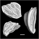
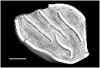
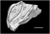
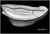
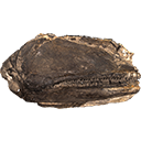
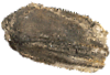
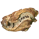
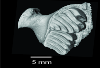
3D models related to the publication: Deciphering the morphological variation and its ontogenetic dynamics in the Late Devonian conodont Icriodus alternatusCatherine Girard, Anne-Lise Charruault
Published online: 08/02/2022 |
|
M3#887conodont element Type: "3D_surfaces"doi: 10.18563/m3.sf.887 state:published |
Download 3D surface file |
Icriodus alternatus UM BUS 032 View specimen
|
M3#888conodont element Type: "3D_surfaces"doi: 10.18563/m3.sf.888 state:published |
Download 3D surface file |
Icriodus alternatus UM BUS 033 View specimen
|
M3#889conodont element Type: "3D_surfaces"doi: 10.18563/m3.sf.889 state:published |
Download 3D surface file |
Icriodus alternatus UM BUS 034 View specimen
|
M3#890conodont element Type: "3D_surfaces"doi: 10.18563/m3.sf.890 state:published |
Download 3D surface file |
Icriodus alternatus UM BUS 035 View specimen
|
M3#891conodont element Type: "3D_surfaces"doi: 10.18563/m3.sf.891 state:published |
Download 3D surface file |
Icriodus alternatus UM BUS 036 View specimen
|
M3#892conodont element Type: "3D_surfaces"doi: 10.18563/m3.sf.892 state:published |
Download 3D surface file |
Icriodus alternatus UM BUS 037 View specimen
|
M3#893conodont element Type: "3D_surfaces"doi: 10.18563/m3.sf.893 state:published |
Download 3D surface file |
Icriodus alternatus UM BUS 038 View specimen
|
M3#894conodont element Type: "3D_surfaces"doi: 10.18563/m3.sf.894 state:published |
Download 3D surface file |
Icriodus alternatus UM BUS 039 View specimen
|
M3#895conodont element Type: "3D_surfaces"doi: 10.18563/m3.sf.895 state:published |
Download 3D surface file |
Icriodus alternatus UM BUS 040 View specimen
|
M3#896conodont element Type: "3D_surfaces"doi: 10.18563/m3.sf.896 state:published |
Download 3D surface file |
Icriodus alternatus UM BUS 041 View specimen
|
M3#897conodont element Type: "3D_surfaces"doi: 10.18563/m3.sf.897 state:published |
Download 3D surface file |
Icriodus alternatus UM BUS 042 View specimen
|
M3#898conodont element Type: "3D_surfaces"doi: 10.18563/m3.sf.898 state:published |
Download 3D surface file |
Icriodus alternatus UM BUS 043 View specimen
|
M3#899conodont element Type: "3D_surfaces"doi: 10.18563/m3.sf.899 state:published |
Download 3D surface file |
Icriodus alternatus UM BUS 044 View specimen
|
M3#900conodont element Type: "3D_surfaces"doi: 10.18563/m3.sf.900 state:published |
Download 3D surface file |
Icriodus alternatus UM BUS 045 View specimen
|
M3#901conodont element Type: "3D_surfaces"doi: 10.18563/m3.sf.901 state:published |
Download 3D surface file |
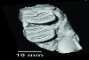
This contribution contains the 3D models of the fossil teeth of two chinchilloid caviomorph rodents (Borikenomys praecursor and Chinchilloidea gen. et sp. indet.) discovered from lower Oligocene deposits of Puerto Rico, San Sebastian Formation (locality LACM Loc. 8060). These fossils were described and figured in the following publication: Marivaux et al. (2020), Early Oligocene chinchilloid caviomorphs from Puerto Rico and the initial rodent colonization of the West Indies. Proceedings of the Royal Society B. http://dx.doi.org/10.1098/rspb.2019.2806
Borikenomys praecursor LACM 162447 View specimen
|
M3#638Right lower m3. This isolated tooth was scanned with a resolution of 6 µm using a μ-CT-scanning station EasyTom 150 / Rx Solutions (Montpellier RIO Imaging, ISE-M, Montpellier, France). AVIZO 7.1 (Visualization Sciences Group) software was used for visualization, segmentation, and 3D rendering. The specimen was prepared within a “labelfield” module of AVIZO, using the segmentation threshold selection tool. Type: "3D_surfaces"doi: 10.18563/m3.sf.638 state:published |
Download 3D surface file |
Borikenomys praecursor LACM 162446 View specimen
|
M3#639Fragment of lower molar (most of the mesial part). This isolated broken tooth was scanned with a resolution of 6 µm using a μ-CT-scanning station EasyTom 150 / Rx Solutions (Montpellier RIO Imaging, ISE-M, Montpellier, France). AVIZO 7.1 (Visualization Sciences Group) software was used for visualization, segmentation, and 3D rendering. The specimen was prepared within a “labelfield” module of AVIZO, using the segmentation threshold selection tool. Type: "3D_surfaces"doi: 10.18563/m3.sf.639 state:published |
Download 3D surface file |
indet indet LACM 162448 View specimen
|
M3#640Fragment of either an upper tooth (mesial laminae) or a lower tooth (distal laminae). The specimen was scanned with a resolution of 6 µm using a μ-CT-scanning station EasyTom 150 / Rx Solutions (Montpellier RIO Imaging, ISE-M, Montpellier, France). AVIZO 7.1 (Visualization Sciences Group) software was used for visualization, segmentation, and 3D rendering. This fragment of tooth was prepared within a “labelfield” module of AVIZO, using the segmentation threshold selection tool. Type: "3D_surfaces"doi: 10.18563/m3.sf.640 state:published |
Download 3D surface file |
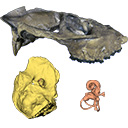
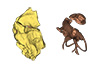
The presented dataset contains the 3D surface scan of the holotype of Birgeria americana, a partial skull described and depicted in: Romano, C., Jenks, J.F., Jattiot, R., Scheyer, T.M., Bylund, K.G. & Bucher, H. 2017. Marine Early Triassic Actinopterygii from Elko County (Nevada, USA): implications for the Smithian equatorial vertebrate eclipse. Journal of Paleontology. https://doi.org/10.1017/jpa.2017.36 .
Birgeria americana NMMNH P-66225 View specimen
|
M3#175NMMNH P-66225 is from upper lower Smithian to lower upper Smithian beds (Thaynes Group). The collecting site is located about 2.75 km south-southeast of the Winecup Ranch, east-central Elko County, Nevada, USA. P-66225 is a partial skull preserved within a large limestone nodule, with its right side exposed. It preserves the portion between the cleithrum posteriorly, and the level of the hind margin of the orbital opening anteriorly. The fossil has a length of 26 cm. Type: "3D_surfaces"doi: 10.18563/m3.sf.175 state:published |
Download 3D surface file |
The present 3D Dataset contains the 3D models analyzed in Hendrickx, C., Gaetano, L. C., Choiniere, J., Mocke, H. and Abdala, F. in press. A new traversodontid cynodont with a peculiar postcanine dentition from the Middle/Late Triassic of Namibia and dental evolution in basal gomphodonts. Journal of Systematic Palaeontology.
Etjoia dentitransitus GSN F1591 View specimen
|
M3#557Surface model derived from µCT data of the holotype of Etjoia dentitransitus Type: "3D_surfaces"doi: 10.18563/m3.sf.557 state:published |
Download 3D surface file |
|
M3#558Photogrammetric 3D surface model of the postcanines of the Holotype of Etjoia dentitransitus Type: "3D_surfaces"doi: 10.18563/m3.sf.558 state:published |
Download 3D surface file |
|
M3#559Photogrammetric 3D surface model of the Holotype of Etjoia dentitransitus Type: "3D_surfaces"doi: 10.18563/m3.sf.559 state:published |
Download 3D surface file |
The present 3D Dataset contains the 3D models analyzed in 3D Finite Element Analysis and Geometric Morphometrics of Sloths (Xenarthra, Folivora) Mandibles Show Insights on the Dietary Specializations of Fossil Taxa. Journal of South American Earth Sciences. https://doi.org/10.1016/j.jsames.2023.104445
Mylodon darwinii CAV 379 View specimen
|
M3#1159Right hemimandible Type: "3D_surfaces"doi: 10.18563/m3.sf.1159 state:published |
Download 3D surface file |
Scelidotherium leptocephalum MNHN-M 137,722 View specimen
|
M3#1160Mandible Type: "3D_surfaces"doi: 10.18563/m3.sf.1160 state:published |
Download 3D surface file |
Glossotherium robustum MNHN-M 914 View specimen
|
M3#1161Mandible Type: "3D_surfaces"doi: 10.18563/m3.sf.1161 state:published |
Download 3D surface file |
Lestodon armatus MPAC 899 View specimen
|
M3#1162Mandible Type: "3D_surfaces"doi: 10.18563/m3.sf.1162 state:published |
Download 3D surface file |
Valgipes bucklandi NHMD.Z.M.K. 1/1845:3540 View specimen
|
M3#1163Mandible Type: "3D_surfaces"doi: 10.18563/m3.sf.1163 state:published |
Download 3D surface file |
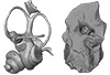
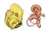
This contribution contains the 3D models of the ossicles of a protocetid archaeocete from the locality of Kpogamé, Togo, described and figured in the publication of Mourlam and Orliac (2019).
indet. indet. UM KPG-M 73 View specimen

|
M3#407stapes Type: "3D_surfaces"doi: 10.18563/m3.sf.407 state:published |
Download 3D surface file |

|
M3#408Incus Type: "3D_surfaces"doi: 10.18563/m3.sf.408 state:published |
Download 3D surface file |

|
M3#409Malleus Type: "3D_surfaces"doi: 10.18563/m3.sf.409 state:published |
Download 3D surface file |
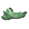
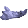
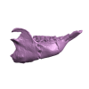
This contribution contains 3D models of extinct rodents Dinomyidae from Miocene and Quaternary of Brazil. The Miocene specimens that were digitalized include the holotypes of Potamarchus adamiae, Pseudopotamarchus villanuevai, and Ferigolomys pacarana collected in the Solimões Formation (Upper Miocene), northern Brazil. The Quaternary specimens are the holotype and paratype of Niedemys piauiensis, found in Upper Pleistocene deposits from northeast Brazil.
Potamarchus adamiae UFAC-CS 011 View specimen
|
M3#410UFAC-CS 011 – holotype, palatal region of the skull with cheek teeth Type: "3D_surfaces"doi: 10.18563/m3.sf.410 state:published |
Download 3D surface file |
Potamarchus adamiae UFAC-CS 043 View specimen
|
M3#411UFAC-CS 043, left dentary with cheek teeth Type: "3D_surfaces"doi: 10.18563/m3.sf.411 state:published |
Download 3D surface file |
Pseudopotamarchus villanuevai UFAC 4762 View specimen
|
M3#412UFAC 4762 – holotype, incomplete right maxilla with cheek teeth Type: "3D_surfaces"doi: 10.18563/m3.sf.412 state:published |
Download 3D surface file |
Ferigolomys pacarana UFAC 6460 View specimen
|
M3#413UFAC 6460 – holotype, palatal region of the skull with cheek teeth Type: "3D_surfaces"doi: 10.18563/m3.sf.413 state:published |
Download 3D surface file |
Drytomomys sp. UFAC 2742 View specimen

|
M3#414UFAC 2742, right dentary with cheek teeth Type: "3D_surfaces"doi: 10.18563/m3.sf.414 state:published |
Download 3D surface file |
Niedemys piauiensis FUMDHAM 113-146365-2 View specimen
|
M3#418FUMDHAM 113-146365-2 - holotype, upper right tooth Type: "3D_surfaces"doi: 10.18563/m3.sf.418 state:published |
Download 3D surface file |
Niedemys piauiensis FUMDHAM 113-145304-2 View specimen
|
M3#419FUMDHAM 113-145304-2 - paratype, left lower molar Type: "3D_surfaces"doi: 10.18563/m3.sf.419 state:published |
Download 3D surface file |
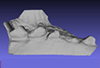
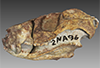
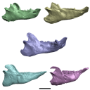
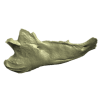
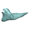
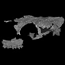
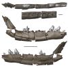
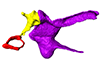
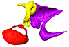
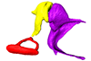
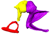
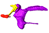
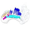
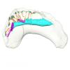
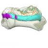
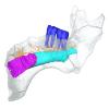
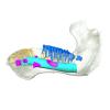
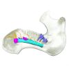
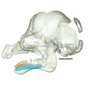
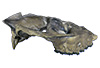
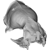
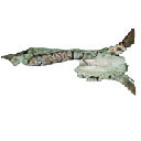
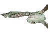
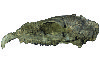
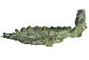
In this work, we digitally restore the snout of the raoellide Khirtharia inflata from the Kalakot area (Rajouri District, Jammu & Kashmir, India). Raoellids are small, semiaquatic ungulates closely related to cetaceans. The specimen is fairly complete and preserves left and right maxillaries, left premaxillary, and part of the anterior and jugal dentition. The digital restoration of this quite complete but deformed specimen of Khirtharia inflata is a welcome addition to the data available for raoellids and will be used to further the understanding of the origins of cetaceans.
Khirtharia inflata GU/RJ/157 View specimen
|
M3#1454deformed partial skull Type: "3D_surfaces"doi: 10.18563/m3.sf.1454 state:published |
Download 3D surface file |
|
M3#1455reconstruction of half snout Type: "3D_surfaces"doi: 10.18563/m3.sf.1455 state:published |
Download 3D surface file |
|
M3#1456reconstruction of complete snout Type: "3D_surfaces"doi: 10.18563/m3.sf.1456 state:published |
Download 3D surface file |
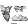
The present 3D Dataset contains the 3D model analyzed in the following publication: Paulina-Carabajal, A., Sterli, J., Werneburg, I., 2019. The endocranial anatomy of the stem turtle Naomichelys speciosa from the Early Cretaceous of North America. Acta Palaeontologica Polonica, https://doi.org/10.4202/app.00606.2019
Naomichelys speciosa FMNH PR273 View specimen

|
M3#428FMNH_PR273_1 - Naomichlys speciosa - skull Type: "3D_surfaces"doi: 10.18563/m3.sf.428 state:published |
Download 3D surface file |
The present 3D Dataset contains the 3D models analyzed in Pochat-Cottilloux Y., Martin J.E., Jouve S., Perrichon G., Adrien J., Salaviale C., de Muizon C., Cespedes R. & Amiot R. (2021). The neuroanatomy of Zulmasuchus querejazus (Crocodylomorpha, Sebecidae) and its implications for the paleoecology of sebecosuchians. The Anatomical Record, https://doi.org/10.1002/ar.24826
Zulmasuchus querejazus MHNC 6672 View specimen

|
M3#798Left endosseous labyrinth of Z. querejazus (MHNC 6672). Type: "3D_surfaces"doi: 10.18563/m3.sf.798 state:published |
Download 3D surface file |

|
M3#799Reconstruction of the endocranial cavities of Z. querejazus (MHNC 6672). Type: "3D_surfaces"doi: 10.18563/m3.sf.799 state:published |
Download 3D surface file |
|
M3#800Three-dimensional reconstruction of the pneumatic cavities within the braincase of Z. querejazus (MHNC 6672) Type: "3D_surfaces"doi: 10.18563/m3.sf.800 state:published |
Download 3D surface file |
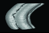
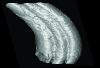
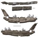
This contribution contains the 3D models described and figured in the following publication: Pujos F., Hautier L., Antoine P-O., Boivin M., Moison B, Salas-Gismondi R, Tejada J.V. , Varas-Malca R.M., Yans J., Marivaux L. (2025). Unexpected pampatheriid from the early Oligocene of Peruvian Amazonia: insights into the tropical differentiation of cingulate xenarthrans. Historical Biology.
Bradypus tridactylus UM-ZOOL-V69 View specimen
|
M3#1600Molariform and associated dentinal microstructure Type: "3D_surfaces"doi: 10.18563/m3.sf.1600 state:published |
Download 3D surface file |
Choloepus didactylus UM-ZOOL-V12 View specimen
|
M3#1601Molariform and associated dentinal microstructure Type: "3D_surfaces"doi: 10.18563/m3.sf.1601 state:published |
Download 3D surface file |
Dasypus mexicanus UM-ZOOL-2787 View specimen
|
M3#1602Molariform and associated dentinal microstructure Type: "3D_surfaces"doi: 10.18563/m3.sf.1602 state:published |
Download 3D surface file |
Tolypeutes matacus UM-ZOOL-2789 View specimen
|
M3#1603Molariform and associated dentinal microstructure Type: "3D_surfaces"doi: 10.18563/m3.sf.1603 state:published |
Download 3D surface file |
Euphractus sexcinctus UM-ZOOL-2790 View specimen
|
M3#1604Molariform and associated dentinal microstructure Type: "3D_surfaces"doi: 10.18563/m3.sf.1604 state:published |
Download 3D surface file |
Holmesina septrionalis UM-FLD-1 View specimen
|
M3#1605Molariform and associated dentinal microstructure Type: "3D_surfaces"doi: 10.18563/m3.sf.1605 state:published |
Download 3D surface file |
Megatherium sp. UM-TAR-1 View specimen
|
M3#1607Molariform and associated dentinal microstructure Type: "3D_surfaces"doi: 10.18563/m3.sf.1607 state:published |
Download 3D surface file |
Indet indet MUSM-3965 View specimen
|
M3#1606Molariform and associated dentinal microstructure Type: "3D_surfaces"doi: 10.18563/m3.sf.1606 state:published |
Download 3D surface file |
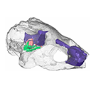
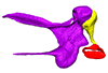
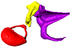
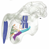
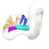
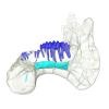
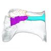
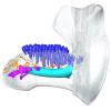
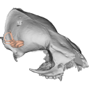
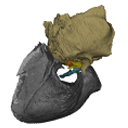
This contribution contains the 3D models described and figured in the following publication: Mennecart B., de Perthuis Ad., Rössner G.E., Guzmán J.A., de Perthuis Au., Costeur L. The first French tragulid skull (Mammalia, Ruminantia, Tragulidae) and associated tragulid remains from the Middle Miocene of Contres (Loir-et-Cher, France). Comptes Rendus Palévol. https://doi.org/10.1016/j.crpv.2017.08.004
Dorcatherium crassum NMB Fa.213.abg View specimen
|
M3#181The 3D surface files of the specimen NMB Fa.213 are the reconstructions of the main skull fragments, the right petrosal bone, and the left bony labyrinth. Type: "3D_surfaces"doi: 10.18563/m3.sf.181 state:published |
Download 3D surface file |
This contribution contains the three-dimensional models of the most informative fossil material attributed to both Peratherium musivum Gernelle, 2024, and Peratherium maximum (Crochet, 1979), respectively from early and middle early Eocene French localities. These specimens, which document the emergence of the relatively large peratheriines, were analyzed and discussed in: Gernelle et al. (2024), Dental morphology evolution in early peratheriines, including a new morphologically cryptic species and findings on the largest early Eocene European metatherian. https://doi.org/10.1080/08912963.2024.2403602
Peratherium musivum MNHN.F.SN122 View specimen
|
M3#16403D surface model of MNHN.F.SN122, right M3 Type: "3D_surfaces"doi: 10.18563/m3.sf.1640 state:published |
Download 3D surface file |
Peratherium musivum MNHN.F.RI220 View specimen
|
M3#16413D surface model of MNHN.F.RI220, left M2 (partial) Type: "3D_surfaces"doi: 10.18563/m3.sf.1641 state:published |
Download 3D surface file |
Peratherium musivum MNHN.F.RI296 View specimen
|
M3#16423D surface model of MNHN.F.RI296, right M1 (partial) Type: "3D_surfaces"doi: 10.18563/m3.sf.1642 state:published |
Download 3D surface file |
Peratherium musivum MNHN.F.RI368 View specimen
|
M3#16433D surface model of MNHN.F.RI368, right m2 Type: "3D_surfaces"doi: 10.18563/m3.sf.1643 state:published |
Download 3D surface file |
Peratherium musivum MNHN.F.RI385 View specimen
|
M3#16443D surface model of MNHN.F.RI385, left m1 Type: "3D_surfaces"doi: 10.18563/m3.sf.1644 state:published |
Download 3D surface file |
Peratherium maximum UM-BRI-17 View specimen
|
M3#16453D surface model of UM-BRI-17, right hemi-mandible with p1-p3, m1-m3 alveoli, and m4 Type: "3D_surfaces"doi: 10.18563/m3.sf.1645 state:published |
Download 3D surface file |
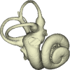
Considerable morphological variations are found in the middle ear among mammals. Here I present a three-dimensional atlas of the middle ear ossicles of eulipotyphlan mammals. This group has radiated into various environments as terrestrial, aquatic, and subterranean habitats independently in multiple lineages. Therefore, eulipotyphlans are an ideal group to explore the form-function relationship of the middle ear ossicles. This comparative atlas of hedgehogs, true shrews, water shrews, mole shrews, true moles, and shrew moles encourages future studies of the middle ear morphology of this diverse group.
Erinaceus europaeus DK2331 View specimen
|
M3#151Left middle ear ossicles Type: "3D_surfaces"doi: 10.18563/m3.sf.151 state:published |
Download 3D surface file |
Anourosorex yamashinai SIK_yamashinai View specimen
|
M3#152Left middle ear ossicles Type: "3D_surfaces"doi: 10.18563/m3.sf.152 state:published |
Download 3D surface file |
Blarina brevicauda M8003 View specimen
|
M3#153Right middle ear ossicles Type: "3D_surfaces"doi: 10.18563/m3.sf.153 state:published |
Download 3D surface file |
Chimarrogale platycephala DK5481 View specimen
|
M3#162Left middle ear ossicles Type: "3D_surfaces"doi: 10.18563/m3.sf.162 state:published |
Download 3D surface file |
Suncus murinus DK1227 View specimen
|
M3#155Left middle ear ossicles Type: "3D_surfaces"doi: 10.18563/m3.sf.155 state:published |
Download 3D surface file |
Condylura cristata SIK0050 View specimen
|
M3#156Right middle ear ossicles Type: "3D_surfaces"doi: 10.18563/m3.sf.156 state:published |
Download 3D surface file |
Euroscaptor klossi SIK0673 View specimen
|
M3#163Left middle ear ossicles Type: "3D_surfaces"doi: 10.18563/m3.sf.163 state:published |
Download 3D surface file |
Euroscaptor malayana SIK_malayana View specimen
|
M3#164Left middle ear ossicles Type: "3D_surfaces"doi: 10.18563/m3.sf.164 state:published |
Download 3D surface file |
Mogera wogura DK2551 View specimen
|
M3#159Left middle ear ossicles Type: "3D_surfaces"doi: 10.18563/m3.sf.159 state:published |
Download 3D surface file |
Talpa altaica SIK_altaica View specimen
|
M3#161Right middle ear ossicles Type: "3D_surfaces"doi: 10.18563/m3.sf.161 state:published |
Download 3D surface file |
Urotrichus talpoides DK0887 View specimen
|
M3#165Left middle ear ossicles Type: "3D_surfaces"doi: 10.18563/m3.sf.165 state:published |
Download 3D surface file |
Oreoscaptor mizura DK6545 View specimen
|
M3#166Left middle ear ossicles Type: "3D_surfaces"doi: 10.18563/m3.sf.166 state:published |
Download 3D surface file |
Scalopus aquaticus SIK_aquaticus View specimen
|
M3#167Left middle ear ossicles Type: "3D_surfaces"doi: 10.18563/m3.sf.167 state:published |
Download 3D surface file |
Scapanus orarius SIK_orarius View specimen
|
M3#168Left middle ear ossicles Type: "3D_surfaces"doi: 10.18563/m3.sf.168 state:published |
Download 3D surface file |
Neurotrichus gibbsii SIK_gibbsii View specimen
|
M3#169Left middle ear ossicles Type: "3D_surfaces"doi: 10.18563/m3.sf.169 state:published |
Download 3D surface file |
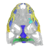
This contribution contains the 3D models of the paratympanic sinus system, the endocast and the neurovascular bony canal of the maxilla, premaxilla and the jugal of Leidyosuchus canadensis and Stangerochampsa mccabei described and figured in the following publication: G. Donzé, G. Perrichon, P. Vincent, JE. Martin, 2026. Comparative endocranial traits in the crocodylians Leidyosuchus canadensis and Stangerochampsa mccabei from the upper Cretaceous of Alberta, Canada. Journal of Anatomy.
Leidyosuchus canadensis TMP1986.221.1 View specimen

|
M3#1830Skull, endocast, paratympanic sinuses, jugal and maxillary neurovascular canals, alveoli Type: "3D_surfaces"doi: 10.18563/m3.sf.1830 state:in_press |
Download 3D surface file |
Stangerochampsa mccabei TMP1986.61.1 View specimen
|
M3#1831Skull, endocast, paratympanic sinuses, jugal and maxillary neurovascular canals, alveoli Type: "3D_surfaces"doi: 10.18563/m3.sf.1831 state:in_press |
Download 3D surface file |
Alligator mississipiensis OUVC 9761 View specimen

|
M3#1833Skull, endocast, paratympanic sinuses, jugal and maxillary neurovascular canals, alveolies Type: "3D_surfaces"doi: 10.18563/m3.sf.1833 state:in_press |
Download 3D surface file |
Alligator sinensis NHMW-Zoo-HS-37966 View specimen

|
M3#1835Skull, endocast, paratympanic sinus, jugal and maxillary neurovascular canals, alveoli Type: "3D_surfaces"doi: 10.18563/m3.sf.1835 state:in_press |
Download 3D surface file |
Crocodylus niloticus MHNL 50001399 View specimen

|
M3#1834Skull, endocast, paratympanic sinuses, jugal and maxillary neurovascular canals, alveolies Type: "3D_surfaces"doi: 10.18563/m3.sf.1834 state:in_press |
Download 3D surface file |
Diplocynodon ratelii LA86 View specimen
|
M3#1832Skull, endocast, paratympanic sinus, jugal and maxillary neurovascular canals, alveolies Type: "3D_surfaces"doi: 10.18563/m3.sf.1832 state:in_press |
Download 3D surface file |

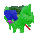
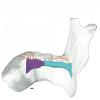
This contribution contains the 3D models described and figured in the following publication: Hautier L, Gomes Rodrigues H, Ferreira-Cardoso S, Emerling CA, Porcher M-L, Asher R, Portela Miguez R, Delsuc F. 2023. From teeth to pad: tooth loss and development of keratinous structures in sirenians. Proceedings of the Royal Society B. https://doi.org/10.1098/rspb.2023.1932
Dugong dugon 2005.51 View specimen
|
M3#1275Internal mandibular morphology. Orange = dorsal canaliculi; purple = mental branches; cyan = mandibular canal; dark blue = teeth; green = tooth alveoli. Type: "3D_surfaces"doi: 10.18563/m3.sf.1275 state:published |
Download 3D surface file |
Dugong dugon 2023.66 View specimen
|
M3#1274Internal mandibular morphology. Orange = dorsal canaliculi; purple = mental branches; cyan = mandibular canal; dark blue = teeth; green = tooth alveoli. Type: "3D_surfaces"doi: 10.18563/m3.sf.1274 state:published |
Download 3D surface file |
Dugong dugon 5386 View specimen
|
M3#1276Internal mandibular morphology. Orange = dorsal canaliculi; purple = mental branches; cyan = mandibular canal; dark blue = teeth; green = tooth alveoli. Type: "3D_surfaces"doi: 10.18563/m3.sf.1276 state:published |
Download 3D surface file |
Dugong dugon 1848.8.29.7/GERM 1027g View specimen
|
M3#1277Internal mandibular morphology. Orange = dorsal canaliculi; purple = mental branches; cyan = mandibular canal; dark blue = teeth. Type: "3D_surfaces"doi: 10.18563/m3.sf.1277 state:published |
Download 3D surface file |
Dugong dugon 1991.413 View specimen
|
M3#1278Internal mandibular morphology. Orange = dorsal canaliculi; purple = mental branches; cyan = mandibular canal; dark blue = teeth. Type: "3D_surfaces"doi: 10.18563/m3.sf.1278 state:published |
Download 3D surface file |
Dugong dugon 1991.427 View specimen
|
M3#1279Internal mandibular morphology. Orange = dorsal canaliculi; purple = mental branches; cyan = mandibular canal; dark blue = teeth. Type: "3D_surfaces"doi: 10.18563/m3.sf.1279 state:published |
Download 3D surface file |
Dugong dugon 2017-3-9 View specimen
|
M3#1280Internal mandibular morphology. Orange = dorsal canaliculi; purple = mental branches; cyan = mandibular canal; dark blue = teeth. Type: "3D_surfaces"doi: 10.18563/m3.sf.1280 state:published |
Download 3D surface file |
Eosiren lybica 1913-22 View specimen
|
M3#1281Internal mandibular morphology. Orange = dorsal canaliculi; purple = mental branches; cyan = mandibular canal; dark blue = teeth; green = tooth alveoli. Type: "3D_surfaces"doi: 10.18563/m3.sf.1281 state:published |
Download 3D surface file |
Halitherium taulannense RGHP C001 View specimen
|
M3#1282Internal mandibular morphology. Cyan = mandibular canal; dark blue = teeth. Type: "3D_surfaces"doi: 10.18563/m3.sf.1282 state:published |
Download 3D surface file |
Halitherium taulannense RGHP C009 View specimen
|
M3#1283Internal mandibular morphology. Orange = dorsal canaliculi; purple = mental branches; cyan = mandibular canal; dark blue = teeth; green = tooth alveoli. Type: "3D_surfaces"doi: 10.18563/m3.sf.1283 state:published |
Download 3D surface file |
Hydrodamalis gigas 1947.10.21.1 View specimen
|
M3#1284Internal mandibular morphology. Orange = dorsal canaliculi; purple = mental branches; cyan = mandibular canal Type: "3D_surfaces"doi: 10.18563/m3.sf.1284 state:published |
Download 3D surface file |
Hydrodamalis gigas C1021 View specimen

|
M3#1285Anterior part of the mandible Type: "3D_surfaces"doi: 10.18563/m3.sf.1285 state:published |
Download 3D surface file |
|
M3#1286Posterior part of the mandible Type: "3D_surfaces"doi: 10.18563/m3.sf.1286 state:published |
Download 3D surface file |
Hydrodamalis gigas 2023.67 View specimen
|
M3#1287Internal mandibular morphology. Orange = dorsal canaliculi; purple = mental branches; cyan = mandibular canal Type: "3D_surfaces"doi: 10.18563/m3.sf.1287 state:published |
Download 3D surface file |
Libysiren sickenbergi M.82429 View specimen
|
M3#1288Internal mandibular morphology. Orange = dorsal canaliculi; purple = mental branches; cyan = mandibular canal; dark blue = teeth; green = tooth alveoli. Type: "3D_surfaces"doi: 10.18563/m3.sf.1288 state:published |
Download 3D surface file |
Libysiren sickenbergi M.45675 View specimen
|
M3#1289Internal mandibular morphology. Orange = dorsal canaliculi; purple = mental branches; cyan = mandibular canal; dark blue = teeth; green = tooth alveoli. Type: "3D_surfaces"doi: 10.18563/m3.sf.1289 state:published |
Download 3D surface file |
Prorastomus sirenoides OR.448976 View specimen

|
M3#1290Internal morphology of the left mandible. Orange = dorsal canaliculi; purple = mental branches; cyan = mandibular canal; dark blue = teeth; green = tooth alveoli. Type: "3D_surfaces"doi: 10.18563/m3.sf.1290 state:published |
Download 3D surface file |
|
M3#1304Internal morphology of the right mandible. Orange = dorsal canaliculi; purple = mental branches; cyan = mandibular canal; dark blue = teeth. Type: "3D_surfaces"doi: 10.18563/m3.sf.1304 state:published |
Download 3D surface file |
Ribodon limbatus M.7073 View specimen
|
M3#1292Internal mandibular morphology. Orange = dorsal canaliculi; purple = mental branches; cyan = mandibular canal; dark blue = teeth; green = tooth alveoli. Type: "3D_surfaces"doi: 10.18563/m3.sf.1292 state:published |
Download 3D surface file |
Rytiodus capgrandi PAL2017-8-1 View specimen
|
M3#1293Internal mandibular morphology. Orange = dorsal canaliculi; purple = mental branches; cyan = mandibular canal; dark blue = teeth; green = tooth alveoli. Type: "3D_surfaces"doi: 10.18563/m3.sf.1293 state:published |
Download 3D surface file |
Trichechus inunguis 1868.12.19.2 View specimen
|
M3#1294Internal mandibular morphology. Orange = dorsal canaliculi; purple = mental branches; cyan = mandibular canal; dark blue = teeth; green = tooth alveoli. Type: "3D_surfaces"doi: 10.18563/m3.sf.1294 state:published |
Download 3D surface file |
Trichechus manatus 1843.3.10.12 View specimen
|
M3#1295Internal mandibular morphology. Orange = dorsal canaliculi; purple = mental branches; cyan = mandibular canal; dark blue = teeth. Type: "3D_surfaces"doi: 10.18563/m3.sf.1295 state:published |
Download 3D surface file |
Trichechus manatus 1864.6.5.1 View specimen
|
M3#1296Internal mandibular morphology. Orange = dorsal canaliculi; purple = mental branches; cyan = mandibular canal; dark blue = teeth; green = tooth alveoli. Type: "3D_surfaces"doi: 10.18563/m3.sf.1296 state:published |
Download 3D surface file |
Trichechus manatus 1950.1.23.1 View specimen
|
M3#1297Internal mandibular morphology. Orange = dorsal canaliculi; purple = mental branches; cyan = mandibular canal; dark blue = teeth; green = tooth alveoli. Type: "3D_surfaces"doi: 10.18563/m3.sf.1297 state:published |
Download 3D surface file |
Trichechus senegalensis 1885.6.30.2 View specimen
|
M3#1298Internal mandibular morphology. Orange = dorsal canaliculi; purple = mental branches; cyan = mandibular canal; dark blue = teeth; green = tooth alveoli. Type: "3D_surfaces"doi: 10.18563/m3.sf.1298 state:published |
Download 3D surface file |
Trichechus senegalensis 1894.7.25.8 View specimen
|
M3#1299Internal mandibular morphology. Orange = dorsal canaliculi; purple = mental branches; cyan = mandibular canal; dark blue = teeth. Type: "3D_surfaces"doi: 10.18563/m3.sf.1299 state:published |
Download 3D surface file |
Trichechus senegalensis V97 View specimen
|
M3#1302Mandibular internal morphology. Orange = dorsal canaliculi; purple = mental branches; cyan = mandibular canal; dark blue = teeth. Type: "3D_surfaces"doi: 10.18563/m3.sf.1302 state:published |
Download 3D surface file |
Trichechus sp. 65.4.28.9 View specimen
|
M3#1300Internal mandibular morphology. Orange = dorsal canaliculi; purple = mental branches; cyan = mandibular canal; dark blue = teeth; green = tooth alveoli. Type: "3D_surfaces"doi: 10.18563/m3.sf.1300 state:published |
Download 3D surface file |
Dugong dugon 1946.8.6.2 View specimen
|
M3#1301Mandibular internal morphology. Orange = dorsal canaliculi; purple = mental branches; cyan = mandibular canal; dark blue = teeth. Type: "3D_surfaces"doi: 10.18563/m3.sf.1301 state:published |
Download 3D surface file |
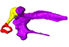
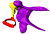
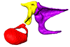
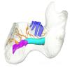
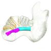
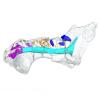
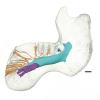
The present 3D Dataset contains the 3D models analyzed in Mennecart B., Métais G., Costeur L., Ginsburg L, and Rössner G. 2021, Reassessment of the enigmatic ruminant Miocene genus Amphimoschus Bourgeois, 1873 (Mammalia, Artiodactyla, Pecora). PlosOne. https://doi.org/10.1371/journal.pone.0244661
Amphimoschus ponteleviensis MNHN.F.AR3266 View specimen
|
M3#701Surface scan of the cast of the skull of Amphimoschus ponteleviensis MNHN.F.AR3266 from Artenay (France) Type: "3D_surfaces"doi: 10.18563/m3.sf.701 state:published |
Download 3D surface file |
|
M3#702Right petrosal bone and bony labyrinth of the skull MNHN.F.AR3266 from Artenay (France) Type: "3D_surfaces"doi: 10.18563/m3.sf.702 state:published |
Download 3D surface file |
Amphimoschus ponteleviensis SMNS40693 View specimen
|
M3#704Left petrosal bone and bony labyrinth of the skull SMNS40693 from Langenau 1 (Germany) Type: "3D_surfaces"doi: 10.18563/m3.sf.704 state:published |
Download 3D surface file |
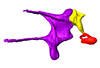
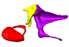
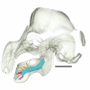
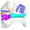
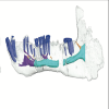
The present 3D Dataset contains 3D models of the cranium surface and of the bony labyrinth endocast of the stem bat Vielasia sigei. They are used by (Hand et al., 2023) to explore the phylogenetic position of this species, to infer its laryngeal echolocating capabilities, and to eventually discuss chiropteran evolution before the crown clade diversification.
Vielasia sigei UM VIE-250 View specimen
|
M3#1269External surface of the cranium Type: "3D_surfaces"doi: 10.18563/m3.sf.1269 state:published |
Download 3D surface file |
|
M3#1270Virtual endocast of the right bony labyrinth Type: "3D_surfaces"doi: 10.18563/m3.sf.1270 state:published |
Download 3D surface file |
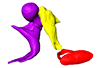
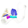
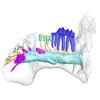
This contribution includes the 3D models of the reconstructed ossicular chain of the cainotheriid Caenomeryx filholi from the late Oligocene locality of Pech Desse (MP28, Quercy, France) described and figured in the publication of Assemat et al. (2020). It represents the oldest ossicular chain reconstruction for a Paleogene terrestrial artiodactyl species.
Caenomeryx filholi UM PDS 3353 View specimen
|
M3#508reconstruction of the middle ear with petrosal, bulla, stapes, incus, malleus Type: "3D_surfaces"doi: 10.18563/m3.sf.508 state:published |
Download 3D surface file |
This contribution contains the 3D models described and figured in: New remains of Neotropical bunodont litopterns and the systematics of Megadolodinae (Mammalia: Litopterna). Geodiversitas.
Megadolodus molariformes VPPLT 974 View specimen
|
M3#1020Partial mandible with the symphysis and left body, bearing the alveoli of ?i2, right and left ?i3, alveolus of right c and p1, roots of left p1, and the left p2–m3 of Megadolodus molariformes (Litopterna, Mammalia) Type: "3D_surfaces"doi: 10.18563/m3.sf.1020 state:published |
Download 3D surface file |
Neodolodus colombianus VPPLT 1696 View specimen
|
M3#1021Almost complete skull with left and right ?I2 and P1–M3 Type: "3D_surfaces"doi: 10.18563/m3.sf.1021 state:published |
Download 3D surface file |
|
M3#1022Partial mandible with complete right and left dentition except for left ?i2 Type: "3D_surfaces"doi: 10.18563/m3.sf.1022 state:published |
Download 3D surface file |


