Explodable 3D Dog Skull for Veterinary Education
3D models of Ocnotherium skull
3D models of Kalakocetus, the earliest Cetacea
3D GM dataset of bird skeletal variation
Skeletal embryonic development in the catshark
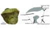
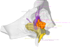
Bony connexions of the petrosal bone of extant hippos
bony labyrinth (14) , inner ear (11) , geometric morphometrics (10) , CT-scan (10) , Eocene (10) , Micro-CT (9) , Miocene (8)
Lionel Hautier (24) , Maëva Judith Orliac (23) , Laurent Marivaux (18) , Renaud Lebrun (14) , Rodolphe Tabuce (14) , Pierre-Olivier Antoine (13) , Bastien Mennecart (13)
|
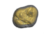
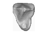
3D models related to the publication: Internal tooth structure and burial practices: insights into the Neolithic necropolis of Gurgy (France, 5100-4000 cal. BC).Mona Le Luyer
Published online: 25/07/2016 |
|
M3#74Outer enamel surface (OES) and enamel-dentine junction (EDJ) of Neolithic upper permanent left second molar Type: "3D_surfaces"doi: 10.18563/m3.sf.74 state:published |
Download 3D surface file |
Homo sapiens GLN04-206-ULM2 View specimen
|
M3#75Outer enamel surface (OES) and enamel-dentine junction (EDJ) of Neolithic upper permanent left second molar Type: "3D_surfaces"doi: 10.18563/m3.sf.75 state:published |
Download 3D surface file |
Homo sapiens GLN05-213-URM2 View specimen
|
M3#76Outer enamel surface (OES) and enamel-dentine junction (EDJ) of Neolithic upper permanent right second molar Type: "3D_surfaces"doi: 10.18563/m3.sf.76 state:published |
Download 3D surface file |
Homo sapiens GLN05-215A-URM2 View specimen
|
M3#77Outer enamel surface (OES) and enamel-dentine junction (EDJ) of Neolithic upper permanent right second molar Type: "3D_surfaces"doi: 10.18563/m3.sf.77 state:published |
Download 3D surface file |
Homo sapiens GLN06-215B-URM2 View specimen
|
M3#78Outer enamel surface (OES) and enamel-dentine junction (EDJ) of Neolithic upper permanent right second molar Type: "3D_surfaces"doi: 10.18563/m3.sf.78 state:published |
Download 3D surface file |
Homo sapiens GLN06-223-URM2 View specimen
|
M3#79Outer enamel surface (OES) and enamel-dentine junction (EDJ) of Neolithic upper permanent right second molar Type: "3D_surfaces"doi: 10.18563/m3.sf.79 state:published |
Download 3D surface file |
Homo sapiens GLN04-229-URM2 View specimen
|
M3#80Outer enamel surface (OES) and enamel-dentine junction (EDJ) of Neolithic upper permanent right second molar Type: "3D_surfaces"doi: 10.18563/m3.sf.80 state:published |
Download 3D surface file |
Homo sapiens GLN05-243B-ULM2 View specimen
|
M3#81Outer enamel surface (OES) and enamel-dentine junction (EDJ) with reconstructed dentine horn tip of Neolithic upper permanent left second molar Type: "3D_surfaces"doi: 10.18563/m3.sf.81 state:published |
Download 3D surface file |
Homo sapiens GLN04-248-ULM2 View specimen
|
M3#82Outer enamel surface (OES) and enamel-dentine junction (EDJ) with reconstructed dentine horn tip of Neolithic upper permanent left second molar Type: "3D_surfaces"doi: 10.18563/m3.sf.82 state:published |
Download 3D surface file |
Homo sapiens GLN04-252-ULM2 View specimen
|
M3#83Outer enamel surface (OES) and enamel-dentine junction (EDJ) of Neolithic upper permanent left second molar Type: "3D_surfaces"doi: 10.18563/m3.sf.83 state:published |
Download 3D surface file |
Homo sapiens GLN04-253-ULM2 View specimen
|
M3#84Outer enamel surface (OES) and enamel-dentine junction (EDJ) of Neolithic upper permanent left second molar Type: "3D_surfaces"doi: 10.18563/m3.sf.84 state:published |
Download 3D surface file |
Homo sapiens GLN05-257-URM2 View specimen
|
M3#85Outer enamel surface (OES) and enamel-dentine junction (EDJ) with reconstructed dentine horn tip of Neolithic upper permanent right second molar Type: "3D_surfaces"doi: 10.18563/m3.sf.85 state:published |
Download 3D surface file |
Homo sapiens GLN04-264-ULM2 View specimen
|
M3#86Outer enamel surface (OES) and enamel-dentine junction (EDJ) of Neolithic upper permanent left second molar Type: "3D_surfaces"doi: 10.18563/m3.sf.86 state:published |
Download 3D surface file |
Homo sapiens GLN04-277-URM2 View specimen
|
M3#87Outer enamel surface (OES) and enamel-dentine junction (EDJ) of Neolithic upper permanent right second molar Type: "3D_surfaces"doi: 10.18563/m3.sf.87 state:published |
Download 3D surface file |
Homo sapiens GLN04-289B-URM2 View specimen
|
M3#88Outer enamel surface (OES) and enamel-dentine junction (EDJ) of Neolithic upper permanent right second molar Type: "3D_surfaces"doi: 10.18563/m3.sf.88 state:published |
Download 3D surface file |
Homo sapiens GLN06-291-URM2 View specimen
|
M3#89Outer enamel surface (OES) and enamel-dentine junction (EDJ) with reconstructed dentine horn tip of Neolithic upper permanent right second molar Type: "3D_surfaces"doi: 10.18563/m3.sf.89 state:published |
Download 3D surface file |
Homo sapiens GLN05-292-URM2 View specimen
|
M3#90Outer enamel surface (OES) and enamel-dentine junction (EDJ) of Neolithic upper permanent right second molar Type: "3D_surfaces"doi: 10.18563/m3.sf.90 state:published |
Download 3D surface file |
Homo sapiens GLN05-294-ULM2 View specimen
|
M3#91Outer enamel surface (OES) and enamel-dentine junction (EDJ) with reconstructed dentine horn tip of Neolithic upper permanent left second molar Type: "3D_surfaces"doi: 10.18563/m3.sf.91 state:published |
Download 3D surface file |
Homo sapiens GLN05-308-URM2 View specimen
|
M3#93Outer enamel surface (OES) and enamel-dentine junction (EDJ) of Neolithic upper permanent right second molar Type: "3D_surfaces"doi: 10.18563/m3.sf.93 state:published |
Download 3D surface file |
Homo sapiens GLN05-301-ULM2 View specimen
|
M3#92Outer enamel surface (OES) and enamel-dentine junction (EDJ) of Neolithic upper permanent left second molar Type: "3D_surfaces"doi: 10.18563/m3.sf.92 state:published |
Download 3D surface file |
This contribution contains the 3D models of the isolated teeth attributed to stem representatives of the Cebuella and Cebus lineages (Cebuella sp. and Cebus sp.), described and figured in the following publication: Marivaux et al. (2016), Dental remains of cebid platyrrhines from the earliest late Miocene of Western Amazonia, Peru: macroevolutionary implications on the extant capuchin and marmoset lineages. American Journal of Physical Anthropology. http://dx.doi.org/10.1002/ajpa.23052
Cebus sp. MUSM-3243 View specimen
|
M3#2823D model of left lower m1 (lingual part) Type: "3D_surfaces"doi: 10.18563/m3.sf.282 state:published |
Download 3D surface file |
Cebuella sp. MUSM-3239 View specimen
|
M3#2833D model of left lower p4 Type: "3D_surfaces"doi: 10.18563/m3.sf.283 state:published |
Download 3D surface file |
Cebuella sp. MUSM-3240 View specimen
|
M3#2943D model of right upper P3 or P4 (buccal part) Type: "3D_surfaces"doi: 10.18563/m3.sf.294 state:published |
Download 3D surface file |
Cebuella sp. MUSM-3241 View specimen
|
M3#2953D model of right upper P2 Type: "3D_surfaces"doi: 10.18563/m3.sf.295 state:published |
Download 3D surface file |
Cebuella sp. MUSM-3242 View specimen
|
M3#2963D model of upper I2 Type: "3D_surfaces"doi: 10.18563/m3.sf.296 state:published |
Download 3D surface file |
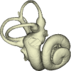
The present 3D Dataset contains 3D models of the cranium surface and of the bony labyrinth endocast of the stem bat Vielasia sigei. They are used by (Hand et al., 2023) to explore the phylogenetic position of this species, to infer its laryngeal echolocating capabilities, and to eventually discuss chiropteran evolution before the crown clade diversification.
Vielasia sigei UM VIE-250 View specimen
|
M3#1269External surface of the cranium Type: "3D_surfaces"doi: 10.18563/m3.sf.1269 state:published |
Download 3D surface file |
|
M3#1270Virtual endocast of the right bony labyrinth Type: "3D_surfaces"doi: 10.18563/m3.sf.1270 state:published |
Download 3D surface file |
This contribution contains the 3D models described and figured in the following publication: Hautier L., Gomes Rodrigues H., Billet G., Asher R.J., 2016. The hidden teeth of sloths: evolutionary vestiges and the development of a simplified dentition. Scientific Reports. doi: 10.1038/srep27763
Bradypus variegatus ZMB 33812 View specimen
|
M3#110Three-dimensional reconstruction of the teeth, mandibles, maxillary and premaxillary bones Type: "3D_surfaces"doi: 10.18563/m3.sf.110 state:published |
Download 3D surface file |
Bradypus variegatus ZMB 41122 View specimen
|
M3#109Three-dimensional reconstruction of the teeth, mandibles, maxillary and premaxillary bones Type: "3D_surfaces"doi: 10.18563/m3.sf.109 state:published |
Download 3D surface file |
Bradypus variegatus MNHN-ZM-MO-1995-326A View specimen
|
M3#111Three-dimensional reconstruction of the teeth, mandibles, maxillary and premaxillary bones Type: "3D_surfaces"doi: 10.18563/m3.sf.111 state:published |
Download 3D surface file |
Bradypus variegatus MNHN-ZM-MO-1995-326B View specimen
|
M3#112Three-dimensional reconstruction of the teeth, mandibles, maxillary and premaxillary bones Type: "3D_surfaces"doi: 10.18563/m3.sf.112 state:published |
Download 3D surface file |
Bradypus sp. MNHN-ZM-MO-1902-325 View specimen
|
M3#113Three-dimensional reconstruction of the teeth, mandibles, maxillary, and premaxillary bones Type: "3D_surfaces"doi: 10.18563/m3.sf.113 state:published |
Download 3D surface file |
Bradypus sp. MNHN-ZM-MO-1995-327 View specimen
|
M3#114Three-dimensional reconstruction of the teeth, mandibles, maxillary and premaxillary bones Type: "3D_surfaces"doi: 10.18563/m3.sf.114 state:published |
Download 3D surface file |
Choloepus didactylus MNHN-ZM-MO-1882-625 View specimen
|
M3#115Three-dimensional reconstruction of the teeth, mandibles, maxillary and premaxillary bones Type: "3D_surfaces"doi: 10.18563/m3.sf.115 state:published |
Download 3D surface file |
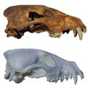
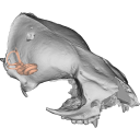
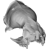
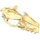
Speothos pacivorus is an extinct South American canid (Canidae: Cerdocyonina) from the Pleistocene of Lagoa Santa Karst, Central Brazil. This taxon is one of the hypercarnivore canids that vanished from the continent at the end of Pleistocene. Although all remains of Speothos pacivorus were collected in the 19th century by the Danish naturalist Peter W. Lund, few studies have committed to an in-depth analysis of the taxon and the known specimens. Here, we analyzed all biological remains of S. pacivorus hosted in the Peter Lund/Quaternary Collection at the Natural History Museum of Denmark, Copenhagen, by listing and illustrating all its specimens known to date. We also conducted a reconstruction of the holotype, an almost complete cranium, based on a µCT scan, producing an undeformed and crack-free three-dimensional model. With this data available we aim to foster new research on this elusive species.
Speothos pacivorus NHMD:211341 View specimen
|
M3#1475Holotype of Speothos pacivorus Type: "3D_surfaces"doi: 10.18563/m3.sf.1475 state:published |
Download 3D surface file |
This contribution includes the 3D models of the reconstructed ossicular chain of the cainotheriid Caenomeryx filholi from the late Oligocene locality of Pech Desse (MP28, Quercy, France) described and figured in the publication of Assemat et al. (2020). It represents the oldest ossicular chain reconstruction for a Paleogene terrestrial artiodactyl species.
Caenomeryx filholi UM PDS 3353 View specimen
|
M3#508reconstruction of the middle ear with petrosal, bulla, stapes, incus, malleus Type: "3D_surfaces"doi: 10.18563/m3.sf.508 state:published |
Download 3D surface file |
The present 3D dataset contains the 3D models of the holotype of Proterochampsa nodosa that were built and analysed in “Redescription, taxonomic revaluation, and phylogenetic affinities of Proterochampsa nodosa (Archosauriformes: Proterochampsidae), early Late Triassic of Candelaria Sequence (Santa Maria Supersequence)”.
Proterochampsa nodosa MCP 1694-PV View specimen
|
M3#9743D models of Proterochampsa nodosa. Model 1: skull. Model 2: mandible. Model 3: left mandibular ramus. Type: "3D_surfaces"doi: 10.18563/m3.sf.974 state:published |
Download 3D surface file |

This contribution contains the 3D models of the fossil remains (maxilla, dentary, and talus) attributed to Djebelemur martinezi, a ca. 50 Ma primate from Tunisia (Djebel Chambi), described and figured in the following publication: Marivaux et al. (2013), Djebelemur, a tiny pre-tooth-combed primate from the Eocene of Tunisia: a glimpse into the origin of crown strepsirhines. PLoS ONE. https://doi.org/10.1371/journal.pone.0080778
Djebelemur martinezi CBI-1-544 View specimen
|
M3#365CBI-1-544, left maxilla preserving P3-M3 and alveoli for P2 and C1 Type: "3D_surfaces"doi: 10.18563/m3.sf.365 state:published |
Download 3D surface file |
Djebelemur martinezi CBI-1-567 View specimen
|
M3#363Isolated left upper P4 Type: "3D_surfaces"doi: 10.18563/m3.sf.363 state:published |
Download 3D surface file |
Djebelemur martinezi CBI-1-565-577-587-580 View specimen
|
M3#366- CBI-1-565, a damaged right mandible, which consists of three isolated pieces found together and reassembled here: the anterior part of the dentary bears the p3 and m1, and alveoli for p4, p2 and c, while the posterior part preserves m3 and a portion of the ascending ramus; the m2 was found isolated but in the same small calcareous block treated by acid processing. - CBI-1-577, isolated right lower p4. - CBI-1-587, isolated left lower p2 (reversed). - CBI-1-580, isolated left lower canine (reversed). Type: "3D_surfaces"doi: 10.18563/m3.sf.366 state:published |
Download 3D surface file |
Djebelemur martinezi CBI-1-545 View specimen
|
M3#364Right Talus Type: "3D_surfaces"doi: 10.18563/m3.sf.364 state:published |
Download 3D surface file |
The present 3D Dataset contains 26 3D models analyzed in the study: On the “cartilaginous rider” in the endocasts of turtle brain cavities, published by the authors in the journal Vertebrate Zoology.
Annemys sp. IVPP-V-18106 View specimen
|
M3#7723D surface(s) file to specimen IVPP-V-18106 Type: "3D_surfaces"doi: 10.18563/m3.sf.772 state:published |
Download 3D surface file |
Apalone spinifera FMNH 22178 View specimen

|
M3#7733D surface(s) file to specimen FMNH 22178 Type: "3D_surfaces"doi: 10.18563/m3.sf.773 state:published |
Download 3D surface file |
Caretta caretta NHMUK1940.3.15.1 View specimen
|
M3#7863D surface(s) file to specimen NHMUK1940.3.15.1 Type: "3D_surfaces"doi: 10.18563/m3.sf.786 state:published |
Download 3D surface file |
Chelodina reimanni ZMB 49659 View specimen

|
M3#7743D surface(s) file to specimen ZMB 49659 Type: "3D_surfaces"doi: 10.18563/m3.sf.774 state:published |
Download 3D surface file |
Chelonia mydas ZMB-37416MS View specimen
|
M3#7753D surface(s) file to specimen ZMB-37416MS Type: "3D_surfaces"doi: 10.18563/m3.sf.775 state:published |
Download 3D surface file |
Cuora amboinensis NHMUK69.42.145_4 View specimen
|
M3#7763D surface(s) file to specimen NHMUK69.42.145_4 Type: "3D_surfaces"doi: 10.18563/m3.sf.776 state:published |
Download 3D surface file |
Emydura subglobosa IW92 View specimen
|
M3#7773D surface(s) file to specimen IW92 Type: "3D_surfaces"doi: 10.18563/m3.sf.777 state:published |
Download 3D surface file |
Eubaena cephalica DMNH 96004 View specimen
|
M3#7783D surface(s) file to specimen DMNH 96004 Type: "3D_surfaces"doi: 10.18563/m3.sf.778 state:published |
Download 3D surface file |
Gopherus berlandieri AMNH-73816 View specimen
|
M3#7793D surface(s) file to specimen AMNH-73816 Type: "3D_surfaces"doi: 10.18563/m3.sf.779 state:published |
Download 3D surface file |
Kinixys belliana AMNH-10028 View specimen
|
M3#7803D surface(s) file to specimen AMNH-10028 Type: "3D_surfaces"doi: 10.18563/m3.sf.780 state:published |
Download 3D surface file |
Macrochelys temminckii GPIT-PV-79430 View specimen
|
M3#7813D surface(s) file to specimen GPIT-PV-79430 Type: "3D_surfaces"doi: 10.18563/m3.sf.781 state:published |
Download 3D surface file |
Malacochersus tornieri SMF-58702 View specimen
|
M3#7873D surface(s) file to specimen SMF-58702 Type: "3D_surfaces"doi: 10.18563/m3.sf.787 state:published |
Download 3D surface file |
Naomichelys speciosa FMNH-PR-273 View specimen
|
M3#7823D surface(s) file to specimen FMNH-PR-273 Type: "3D_surfaces"doi: 10.18563/m3.sf.782 state:published |
Download 3D surface file |
Pelodiscus sinensis IW576-2 View specimen
|
M3#7833D surface(s) file to specimen IW576-2 Type: "3D_surfaces"doi: 10.18563/m3.sf.783 state:published |
Download 3D surface file |
Platysternon megacephalum SMF-69684 View specimen
|
M3#7843D surface(s) file to specimen SMF-69684 Type: "3D_surfaces"doi: 10.18563/m3.sf.784 state:published |
Download 3D surface file |
Podocnemis unifilis SMF-55470 View specimen
|
M3#7853D surface(s) file to specimen SMF-55470 Type: "3D_surfaces"doi: 10.18563/m3.sf.785 state:published |
Download 3D surface file |
Proganochelys quenstedtii MB 1910.45.2 View specimen
|
M3#7883D surface(s) file to specimen MB 1910.45.2 Type: "3D_surfaces"doi: 10.18563/m3.sf.788 state:published |
Download 3D surface file |
Proganochelys quenstedtii SMNS 16980 View specimen
|
M3#7893D surface(s) file to specimen SMNS 16980 Type: "3D_surfaces"doi: 10.18563/m3.sf.789 state:published |
Download 3D surface file |
Rhinochelys pulchriceps CAMSM_B55775 View specimen
|
M3#7903D surface(s) file to specimen CAMSM_B55775 Type: "3D_surfaces"doi: 10.18563/m3.sf.790 state:published |
Download 3D surface file |
Rhinoclemmys funereal YPM12174 View specimen
|
M3#7913D surface(s) file to specimen YPM12174 Type: "3D_surfaces"doi: 10.18563/m3.sf.791 state:published |
Download 3D surface file |
Sandownia harrisi MIWG3480 View specimen
|
M3#7923D surface(s) file to specimen MIWG3480 Type: "3D_surfaces"doi: 10.18563/m3.sf.792 state:published |
Download 3D surface file |
Testudo graeca YPM14342 View specimen
|
M3#7933D surface(s) file to specimen YPM14342 Type: "3D_surfaces"doi: 10.18563/m3.sf.793 state:published |
Download 3D surface file |
Testudo hermanni AMNH134518 View specimen
|
M3#7943D surface(s) file to specimen AMNH134518 Type: "3D_surfaces"doi: 10.18563/m3.sf.794 state:published |
Download 3D surface file |
Trachemys scripta NN View specimen
|
M3#7953D surface(s) file to specimen Trachemys scripta Type: "3D_surfaces"doi: 10.18563/m3.sf.795 state:published |
Download 3D surface file |
Xinjiangchelys radiplicatoides IVPP V9539 View specimen
|
M3#7963D surface(s) file to specimen IVPP V9539 Type: "3D_surfaces"doi: 10.18563/m3.sf.796 state:published |
Download 3D surface file |
Chelydra serpentina UFR VP1 View specimen

|
M3#801Brain endocast Type: "3D_surfaces"doi: 10.18563/m3.sf.801 state:published |
Download 3D surface file |
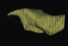
The present 3D Dataset contains the 3D models of the endocranial cast of two specimens of Indohyus indirae described in the article entitled “The endocranial cast of Indohyus (Artiodactyla, Raoellidae): the origin of the cetacean brain” (Orliac and Thewissen, 2021). They represent the cast of the main cavity of the braincase as well as associated intraosseous sinuses.
Indohyus indirae RR 207 View specimen
|
M3#710cast of the main endocranial cavity and associated intraosseous sinuses Type: "3D_surfaces"doi: 10.18563/m3.sf.710 state:published |
Download 3D surface file |
Indohyus indirae RR 601 View specimen
|
M3#711casts of the main endocranial cavity Type: "3D_surfaces"doi: 10.18563/m3.sf.711 state:published |
Download 3D surface file |
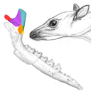
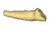
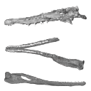
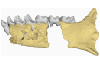
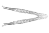
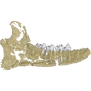
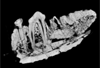
This note presents the 3D model of the hemi-mandible UM-PAT 159 of the MP7 Diacodexis species D. cf. gigasei and 3D models corresponding to the restoration of the ascending ramus, broken on the original specimen, and to a restoration of a complete mandible based on the preserved left hemi-mandible.
Diacodexis cf. gigasei UMPAT159 View specimen
|
M3#3153D models of UM PAT 159 after the restoration of the ascending ramus Type: "3D_surfaces"doi: 10.18563/m3.sf.315 state:published |
Download 3D surface file |
|
M3#316restoration of a complete mandible based on the preserved left hemi-mandible UM PAT 159 Type: "3D_surfaces"doi: 10.18563/m3.sf.316 state:published |
Download 3D surface file |
|
M3#3173D model of the hemi-mandible UM PAT 159 Type: "3D_surfaces"doi: 10.18563/m3.sf.317 state:published |
Download 3D surface file |
This contribution contains the 3D models described and figured in: The Neogene record of northern South American native ungulates. Smithsonian Contributions to Paleobiology. Doi: 10.5479/si.1943-6688.101
Hilarcotherium miyou IGMp 881327 View specimen
|
M3#318Right upper M2 Type: "3D_surfaces"doi: 10.18563/m3.sf.318 state:published |
Download 3D surface file |
Hilarcotherium miyou MUN-STRI 34216 View specimen
|
M3#319Right upper P4 Type: "3D_surfaces"doi: 10.18563/m3.sf.319 state:published |
Download 3D surface file |
|
M3#320Right upper M2 Type: "3D_surfaces"doi: 10.18563/m3.sf.320 state:published |
Download 3D surface file |
Falcontoxodon aguilerai AMU-CURS 585 View specimen
|
M3#321Maxilla with left M3-P2 and right I2 Type: "3D_surfaces"doi: 10.18563/m3.sf.321 state:published |
Download 3D surface file |
This project presents a µCT dataset and an associated 3D surface model of the holotype of Donrussellia magna (UM PAT 17; Primates, Adapiformes). UM PAT17 is the only known specimen for the species and consists of a well-preserved left lower jaw with p4-m3. It documents one of the oldest European primates, eventually dated near the Paleocene Eocene Thermal Maximum.
Donrussellia magna UM PAT 17 View specimen

|
M3#173D surface file model of UM PAT 17 (type specimen of Donrussellia magna), which is a well preserved left lower jaw with p4-m3. The teeth (and roots) were manually segmented. Type: "3D_surfaces"doi: 10.18563/m3.sf17 state:published |
Download 3D surface file |
|
M3#18CT Scan Data of Donrussellia magna UM PAT 17. Voxel size (in µm): 36µm (isotropic voxels). Dimensions in x,y,z : 594 pixels, 294 pixels, 1038 pixels. Image type : 8-bit voxels. Image format : raw data format (no header). Type: "3D_CT"doi: 10.18563/m3.sf18 state:published |
Download CT data |
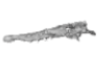
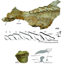
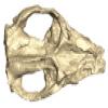
This contribution contains the 3D models described and figured in the following publication: Kassegne K. E., Mourlam M. J., Guinot G., Amoudji Y. Z., Martin J. E., Togbe K. A., Johnson A. K., Hautier L. 2021. First partial cranium of Togocetus from Kpogamé (Togo) and the protocetid diversity in the Togolese phosphate basin. Annales de Paléontologie, Issue 2, April–June 2021, 102488. https://doi.org/10.1016/j.annpal.2021.102488
Togocetus cf. traversei ULDG-KPO1 View specimen
|
M3#768The specimen consists of a partial cranium prepared out of a calcareous phosphate matrix. The partial cranium lacks the anterior part of the rostrum, the cranial roof, and most of the basicranium apart from the left zygomatic process of the squamosal. The maxilla, nasal, palatine, pterygoid, alisphenoid, and squamosal bones are preserved, as well as two incomplete dental rows described hereafter. Type: "3D_surfaces"doi: 10.18563/m3.sf.768 state:published |
Download 3D surface file |
|
M3#770µCT . Resolution: 0.3156mm. This scan can easily be opened with Fiji, MorphoDig, 3DSlicer, or any software that reads .MHD file format. Also, the .RAW file can be opened easily with other software such as Avizo/Amira when providing the correct dimensions (which are enclosed within the file name) Type: "3D_CT"doi: 10.18563/m3.sf.770 state:published |
Download CT data |
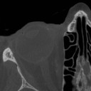
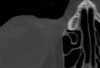
The present Dataset contains the micro-CT scan of the head of an anonymous 54 year old female donor, at a voxel resolution of 145µm. The skin of the face has been masked in order to avoid the donor to be recognized.
Homo sapiens UM_HS_2018_09_13 View specimen
|
M3#1152Micro-ct data set Type: "3D_CT"doi: 10.18563/m3.sf.1152 state:published |
Download CT data |
The present 3D Dataset contains the 3D models of the sacral vertebrae analyzed in “Sacral co-ossification in dinosaurs: The oldest record of fused sacral vertebrae in Dinosauria and the diversity of sacral co-ossification patterns in the group”.
Buriolestes schultzi CAPPA/UFSM 0035 View specimen
|
M3#705Sacral vertebrae of Buriolestes schultzi Type: "3D_surfaces"doi: 10.18563/m3.sf.705 state:published |
Download 3D surface file |
indet indet CAPPA/UFSM 0228 View specimen
|
M3#706Sacral vertebrae of a saurischian dinosaur indet. Type: "3D_surfaces"doi: 10.18563/m3.sf.706 state:published |
Download 3D surface file |
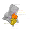
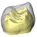
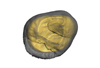
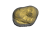
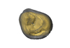
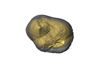
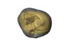
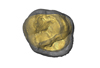
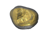
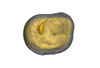
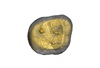
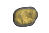
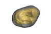
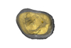
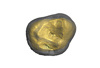
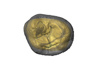
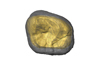
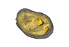
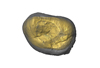
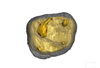
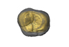
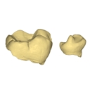
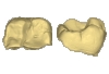
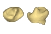
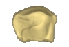
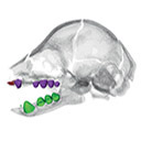
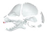
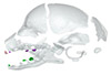
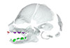
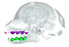
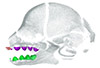
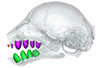
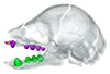
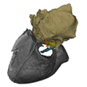
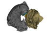
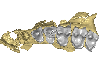
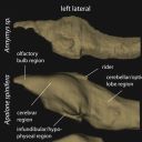
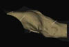
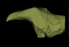
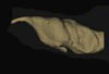
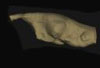
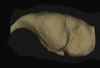
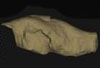
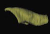
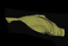
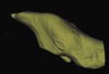
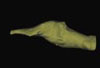
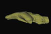
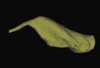
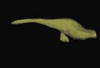
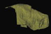
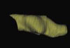
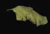
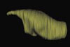
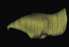
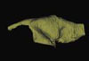
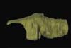
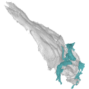
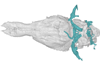
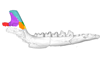
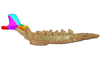
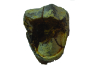
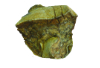
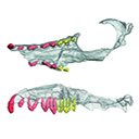
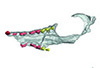
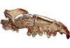
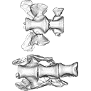
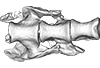
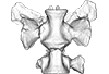
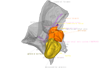
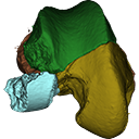
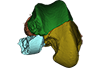
This project presents the osteological connexions of the petrosal bone of the extant Hippopotamidae Hippopotamus amphibius and Choeropsis liberiensis by a virtual osteological dissection of the ear region. The petrosal, the bulla, the sinuses and the major morphological features surrounding the petrosal bone are labelled, both in situ and in an exploded model presenting disassembly views. The directional underwater hearing mode of Hippopotamidae is discussed based on the new observations.
Choeropsis liberiensis UPPal-M09-5-005a View specimen
|
M3#1Labelled compact model of the right ear region of Choeropsis liberiensis (UPPal-M09-5-005a) Type: "3D_surfaces"doi: 10.18563/m3.sf1 state:published |
Download 3D surface file |

|
M3#2Labelled exploded model of the right ear region of Choeropsis liberiensis (UPPal-M09-5-005a) Type: "3D_surfaces"doi: 10.18563/m3.sf2 state:published |
Download 3D surface file |
Hippopotamus amphibius UM N179 View specimen
|
M3#3Labelled compact model of the right ear region of Hippopotamus amphibius (UM N 179) Type: "3D_surfaces"doi: 10.18563/m3.sf3 state:published |
Download 3D surface file |

|
M3#4Labelled exploded model of the right ear region of Hippopotamus amphibius (UM N 179) Type: "3D_surfaces"doi: 10.18563/m3.sf4 state:published |
Download 3D surface file |
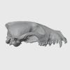
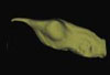
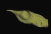
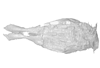
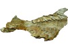
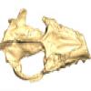
The present 3D Dataset contains the 3D model of the skull of the raoellid Indohyus indirae described in Patel et al. 2024.
Indohyus indirae RR 207 View specimen
|
M3#1259dorsoventrally crushed skull Type: "3D_surfaces"doi: 10.18563/m3.sf.1259 state:published |
Download 3D surface file |
Indohyus indirae RR 601 View specimen
|
M3#1268dorsoventrally crushed skull Type: "3D_surfaces"doi: 10.18563/m3.sf.1268 state:published |
Download 3D surface file |
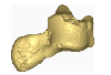
The present contribution contains the 3D virtual restoration of a Pliocene Lutrine right femur of Tobène, Senegal, described and figured in Lihoreau et al. (2021) : "A fossil terrestrial fauna from Tobène (Senegal) provides a unique early Pliocene window in Western Africa ". https://doi.org/10.1016/j.gr.2021.06.013
Indet indet SN-Tob-12-02 View specimen
|
M3#441Virtual restoration of SN-Tob-12-02 Type: "3D_surfaces"doi: 10.18563/m3.sf.441 state:published |
Download 3D surface file |
This contribution contains the 3D models described and figured in the following publication: Paulina-Carabajal, A. and Nieto, M. N. In press. Brief comment on the brain and inner ear of Giganotosaurus carolinii (Dinosauria: Theropoda) based on CT scans. Ameghiniana. https://doi.org/10.5710/AMGH.25.10.2019.3237
Giganotosaurus carolinii MUCPv-CH-1 View specimen
|
M3#504The current file contents 3D models of the braincase, brain, left and right inner ears Type: "3D_surfaces"doi: 10.18563/m3.sf.504 state:published |
Download 3D surface file |


