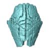
Explodable 3D Dog Skull for Veterinary Education
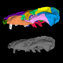
3D models of Ocnotherium skull
3D models of Kalakocetus, the earliest Cetacea
3D GM dataset of bird skeletal variation
Skeletal embryonic development in the catshark
Bony connexions of the petrosal bone of extant hippos
bony labyrinth (14) , inner ear (11) , geometric morphometrics (10) , CT-scan (10) , Eocene (10) , Micro-CT (9) , Miocene (8)
Lionel Hautier (24) , Maëva Judith Orliac (23) , Laurent Marivaux (18) , Renaud Lebrun (14) , Rodolphe Tabuce (14) , Pierre-Olivier Antoine (13) , Bastien Mennecart (13)
|
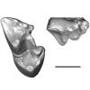
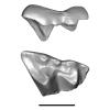
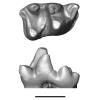
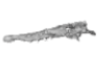
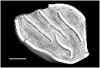
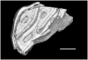
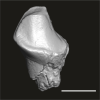
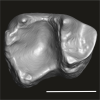
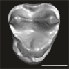
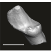
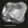
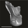
A mandible of Diacodexis cf. gigasei (Artiodactyla, Diacodexeidae) from the Early Eocene locality of Palette (Bouches-du-Rhône, France)Maëva J. Orliac
Published online: 03/07/2018 |
|
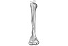
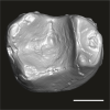
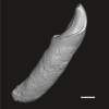
M3#3153D models of UM PAT 159 after the restoration of the ascending ramus Type: "3D_surfaces"doi: 10.18563/m3.sf.315 state:published |
Download 3D surface file |
|
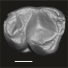
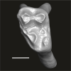
M3#316restoration of a complete mandible based on the preserved left hemi-mandible UM PAT 159 Type: "3D_surfaces"doi: 10.18563/m3.sf.316 state:published |
Download 3D surface file |
|
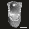
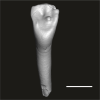
M3#3173D model of the hemi-mandible UM PAT 159 Type: "3D_surfaces"doi: 10.18563/m3.sf.317 state:published |
Download 3D surface file |
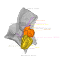
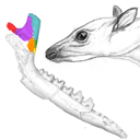
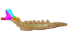
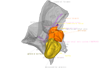
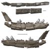
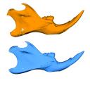
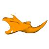
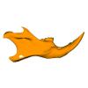
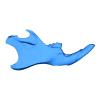
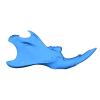
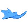
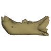
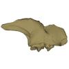
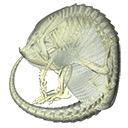
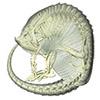
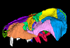
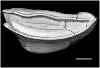
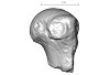
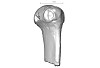
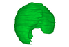
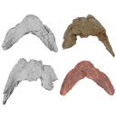
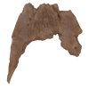
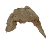
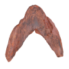
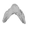
This project presents the osteological connexions of the petrosal bone of the extant Hippopotamidae Hippopotamus amphibius and Choeropsis liberiensis by a virtual osteological dissection of the ear region. The petrosal, the bulla, the sinuses and the major morphological features surrounding the petrosal bone are labelled, both in situ and in an exploded model presenting disassembly views. The directional underwater hearing mode of Hippopotamidae is discussed based on the new observations.
Choeropsis liberiensis UPPal-M09-5-005a View specimen
|
M3#1Labelled compact model of the right ear region of Choeropsis liberiensis (UPPal-M09-5-005a) Type: "3D_surfaces"doi: 10.18563/m3.sf1 state:published |
Download 3D surface file |

|
M3#2Labelled exploded model of the right ear region of Choeropsis liberiensis (UPPal-M09-5-005a) Type: "3D_surfaces"doi: 10.18563/m3.sf2 state:published |
Download 3D surface file |
Hippopotamus amphibius UM N179 View specimen

|
M3#3Labelled compact model of the right ear region of Hippopotamus amphibius (UM N 179) Type: "3D_surfaces"doi: 10.18563/m3.sf3 state:published |
Download 3D surface file |

|
M3#4Labelled exploded model of the right ear region of Hippopotamus amphibius (UM N 179) Type: "3D_surfaces"doi: 10.18563/m3.sf4 state:published |
Download 3D surface file |
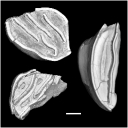
This contribution contains the three-dimensional models of the most informative fossil material attributed to both Peratherium musivum Gernelle, 2024, and Peratherium maximum (Crochet, 1979), respectively from early and middle early Eocene French localities. These specimens, which document the emergence of the relatively large peratheriines, were analyzed and discussed in: Gernelle et al. (2024), Dental morphology evolution in early peratheriines, including a new morphologically cryptic species and findings on the largest early Eocene European metatherian. https://doi.org/10.1080/08912963.2024.2403602
Peratherium musivum MNHN.F.SN122 View specimen
|
M3#16403D surface model of MNHN.F.SN122, right M3 Type: "3D_surfaces"doi: 10.18563/m3.sf.1640 state:published |
Download 3D surface file |
Peratherium musivum MNHN.F.RI220 View specimen
|
M3#16413D surface model of MNHN.F.RI220, left M2 (partial) Type: "3D_surfaces"doi: 10.18563/m3.sf.1641 state:published |
Download 3D surface file |
Peratherium musivum MNHN.F.RI296 View specimen
|
M3#16423D surface model of MNHN.F.RI296, right M1 (partial) Type: "3D_surfaces"doi: 10.18563/m3.sf.1642 state:published |
Download 3D surface file |
Peratherium musivum MNHN.F.RI368 View specimen
|
M3#16433D surface model of MNHN.F.RI368, right m2 Type: "3D_surfaces"doi: 10.18563/m3.sf.1643 state:published |
Download 3D surface file |
Peratherium musivum MNHN.F.RI385 View specimen
|
M3#16443D surface model of MNHN.F.RI385, left m1 Type: "3D_surfaces"doi: 10.18563/m3.sf.1644 state:published |
Download 3D surface file |
Peratherium maximum UM-BRI-17 View specimen
|
M3#16453D surface model of UM-BRI-17, right hemi-mandible with p1-p3, m1-m3 alveoli, and m4 Type: "3D_surfaces"doi: 10.18563/m3.sf.1645 state:published |
Download 3D surface file |
This contribution contains 3D models of mandibles of Cypriot mice (Mus cypriacus) and house mice (Mus musculus domesticus) from the island of Cyprus. The niche partitioning of the two species was investigated using isotopic ecology, geometric morphometrics and biomechanics. Both species displayed generalist feeding behavior, modulated by fine-tuned adaptation to their feeding habits. The house mouse mandible, with a relatively large masseter area and an optimization for incisor biting, appears as an all-rounder tool for foraging on diverse non-natural items.
These models are analyzed in the following publication: Renaud et al 2024, “Trophic differentiation between the endemic Cypriot mouse and the house mouse: a study coupling stable isotopes and morphometrics”, https://doi.org/10.1007/s10914-024-09740-5
Mus cypriacus Cypriacus_5GE View specimen
|
M3#15843D model of the right mandible Type: "3D_surfaces"doi: 10.18563/m3.sf.1584 state:published |
Download 3D surface file |
Mus cypriacus Cypriacus_BET2 View specimen
|
M3#15853D model of the right mandible Type: "3D_surfaces"doi: 10.18563/m3.sf.1585 state:published |
Download 3D surface file |
Mus cypriacus Cypriacus_FON1 View specimen
|
M3#15863D model of the right mandible Type: "3D_surfaces"doi: 10.18563/m3.sf.1586 state:published |
Download 3D surface file |
Mus cypriacus Cypriacus_FON2 View specimen

|
M3#15873D model of the right mandible Type: "3D_surfaces"doi: 10.18563/m3.sf.1587 state:published |
Download 3D surface file |
Mus cypriacus Cypriacus_KOU1 View specimen
|
M3#15883D model of the right mandible Type: "3D_surfaces"doi: 10.18563/m3.sf.1588 state:published |
Download 3D surface file |
Mus musculus Cyprus_dom_KOF1 View specimen
|
M3#15893D model of the right mandible Type: "3D_surfaces"doi: 10.18563/m3.sf.1589 state:published |
Download 3D surface file |
Mus musculus Cyprus_dom_LEF1 View specimen
|
M3#15903D model of the right mandible Type: "3D_surfaces"doi: 10.18563/m3.sf.1590 state:published |
Download 3D surface file |
Mus musculus Cyprus_dom_MEN1 View specimen

|
M3#15913D model of the right mandible Type: "3D_surfaces"doi: 10.18563/m3.sf.1591 state:published |
Download 3D surface file |
Mus musculus Cyprus_dom_TSE2 View specimen
|
M3#15923D model of the mirrored left mandible Type: "3D_surfaces"doi: 10.18563/m3.sf.1592 state:published |
Download 3D surface file |
Mus musculus Cyprus_dom_XYL5 View specimen
|
M3#15933D model of the right mandible Type: "3D_surfaces"doi: 10.18563/m3.sf.1593 state:published |
Download 3D surface file |
This contribution contains the 3D model described and figured in the following publication: Martin, T., Averianov, A. O., Schultz, J. A., & Schwermann, A. H. (2023). A stem therian mammal from the Lower Cretaceous of Germany. Journal of Vertebrate Paleontology, e2224848.
Spelaeomolitor speratus WMNM P99101 View specimen

|
M3#12573D_model_Spelaeomolitor_lower_molar Type: "3D_surfaces"doi: 10.18563/m3.sf.1257 state:published |
Download 3D surface file |

|
M3#1258CT imagestack (jpgs) and info data sheet (pca file) in one zip folder Type: "3D_CT"doi: 10.18563/m3.sf.1258 state:published |
Download CT data |
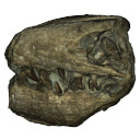
This contribution contains the 3D models described and figured in the following publication: Bonis et al. 2023. A new large pantherine and a sabre-toothed cat (Mammalia, Carnivora, Felidae) from the late Miocene hominoid-bearing Khorat sand pits, Nakhon Ratchasima Province, northeastern Thailand. The Science of Nature 110(5):42. https://doi.org/10.1007/s00114-023-01867-4
Pachypanthera piriyai CUF-KR-1 View specimen
|
M3#1209Holotype of Pachypanthera piriyai, a left hemi-mandible with alveoli for i1-i3 and canine, roots of p3, p4 and partially broken off m1 crown. Type: "3D_surfaces"doi: 10.18563/m3.sf.1209 state:published |
Download 3D surface file |
Pachypanthera piriyai CUF-KR-2 View specimen
|
M3#1210Paratype of Pachypanthera piriyai, a right hemi-maxilla with P3-P4, alveoli of C and M1, root of P2 Type: "3D_surfaces"doi: 10.18563/m3.sf.1210 state:published |
Download 3D surface file |
This contribution contains 3D models of the holotype of a new species of long-nosed armadillos, the Guianan long-nosed armadillo (Dasypus guianensis) described in the following publication: Barthe M., Rancilhac L., Arteaga M. C., Feijó A., Tilak M.-K., Justy F., Loughry W. J., McDonough C. M., de Thoisy B., Catzeflis F., Billet G., Hautier L., Nabholz B., and Delsuc F. 2024. Exon capture museomics deciphers the nine-banded armadillo species complex and identifies a new species endemic to the Guiana Shield. Systematic Biology, syae027. https://doi.org/10.1093/sysbio/syae027
Dasypus guianensis MNHN-ZM-MO-2001-1317 View specimen
|
M3#1200Skeleton and carapace Type: "3D_surfaces"doi: 10.18563/m3.sf.1200 state:published |
Download 3D surface file |
|
M3#1201Frontal sinuses Type: "3D_surfaces"doi: 10.18563/m3.sf.1201 state:published |
Download 3D surface file |
This contribution contains the three-dimensional models of the inner ear of the hetaxodontid rodents Amblyrhiza, Clidomys and Elasmodontomys from the West Indies. These specimens were analyzed and discussed in : The inner ear of caviomorph rodents: phylogenetic implications and application to extinct West Indian taxa.
Amblyrhiza inundata 11842 View specimen

|
M3#11543D surface of the left-oriented inner ear of Amblyrhiza. Type: "3D_surfaces"doi: 10.18563/m3.sf.1154 state:published |
Download 3D surface file |
Clidomys sp NA View specimen
|
M3#11553D surface of the left-oriented inner ear of Clidomys sp. Type: "3D_surfaces"doi: 10.18563/m3.sf.1155 state:published |
Download 3D surface file |
Elasmodontomys obliquus 17127 View specimen

|
M3#11563D surface of the left-oriented inner ear of Elasmodontomys obliquus. Type: "3D_surfaces"doi: 10.18563/m3.sf.1156 state:published |
Download 3D surface file |
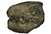
The present 3D Dataset contains the 3D model analyzed in the following publication: Carolina A. Hoffmann, A. G. Martinelli & M. B. Andrade. 2023. Anatomy of the holotype of “Probelesodon” kitchingi revisited, a chiniquodontid cynodont (Synapsida, Probainognathia) from the early Late Triassic of southern Brazil, Journal of Paleontology
Probelesodon kitchingi MCP 1600 PV View specimen
|
M3#11513D models of the skull with segmented bones and without the segmentation. colormap and orientation files also added. Type: "3D_surfaces"doi: 10.18563/m3.sf.1151 state:published |
Download 3D surface file |
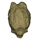
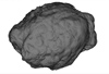
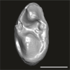
The present 3D Dataset contains the 3D model of the endocranial cast of Palaeolama sp. from the mid-Pleistocene (~1.2 Mya) of South America, analyzed in Balcarcel et al. 2023.
Palaeolama sp. PIMUZ A/V 4091 View specimen
|
M3#11283D model of a natural endocast Type: "3D_surfaces"doi: 10.18563/m3.sf.1128 state:published |
Download 3D surface file |
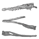
The present 3D dataset contains the 3D models of the holotype of Proterochampsa nodosa that were built and analysed in “Redescription, taxonomic revaluation, and phylogenetic affinities of Proterochampsa nodosa (Archosauriformes: Proterochampsidae), early Late Triassic of Candelaria Sequence (Santa Maria Supersequence)”.
Proterochampsa nodosa MCP 1694-PV View specimen
|
M3#9743D models of Proterochampsa nodosa. Model 1: skull. Model 2: mandible. Model 3: left mandibular ramus. Type: "3D_surfaces"doi: 10.18563/m3.sf.974 state:published |
Download 3D surface file |
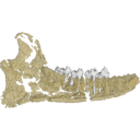
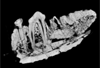
This contribution contains the 3D models of the fossil teeth of two chinchilloid caviomorph rodents (Borikenomys praecursor and Chinchilloidea gen. et sp. indet.) discovered from lower Oligocene deposits of Puerto Rico, San Sebastian Formation (locality LACM Loc. 8060). These fossils were described and figured in the following publication: Marivaux et al. (2020), Early Oligocene chinchilloid caviomorphs from Puerto Rico and the initial rodent colonization of the West Indies. Proceedings of the Royal Society B. http://dx.doi.org/10.1098/rspb.2019.2806
Borikenomys praecursor LACM 162447 View specimen
|
M3#638Right lower m3. This isolated tooth was scanned with a resolution of 6 µm using a μ-CT-scanning station EasyTom 150 / Rx Solutions (Montpellier RIO Imaging, ISE-M, Montpellier, France). AVIZO 7.1 (Visualization Sciences Group) software was used for visualization, segmentation, and 3D rendering. The specimen was prepared within a “labelfield” module of AVIZO, using the segmentation threshold selection tool. Type: "3D_surfaces"doi: 10.18563/m3.sf.638 state:published |
Download 3D surface file |
Borikenomys praecursor LACM 162446 View specimen
|
M3#639Fragment of lower molar (most of the mesial part). This isolated broken tooth was scanned with a resolution of 6 µm using a μ-CT-scanning station EasyTom 150 / Rx Solutions (Montpellier RIO Imaging, ISE-M, Montpellier, France). AVIZO 7.1 (Visualization Sciences Group) software was used for visualization, segmentation, and 3D rendering. The specimen was prepared within a “labelfield” module of AVIZO, using the segmentation threshold selection tool. Type: "3D_surfaces"doi: 10.18563/m3.sf.639 state:published |
Download 3D surface file |
indet indet LACM 162448 View specimen
|
M3#640Fragment of either an upper tooth (mesial laminae) or a lower tooth (distal laminae). The specimen was scanned with a resolution of 6 µm using a μ-CT-scanning station EasyTom 150 / Rx Solutions (Montpellier RIO Imaging, ISE-M, Montpellier, France). AVIZO 7.1 (Visualization Sciences Group) software was used for visualization, segmentation, and 3D rendering. This fragment of tooth was prepared within a “labelfield” module of AVIZO, using the segmentation threshold selection tool. Type: "3D_surfaces"doi: 10.18563/m3.sf.640 state:published |
Download 3D surface file |
The present 3D Dataset contains the 3D model used in in the following publication: Interacting with the inaccessible: utilization of multimedia-based visual contents of Japan’s National Monument, the Taniwhasaurus mikasaensis (Mosasauridae) holotype for educational workshops at Mikasa City Museum.
Taniwhasaurus mikasaensis MCM.M0009 View specimen
|
M3#499Taniwhasaurus mikasaensis, Caldwell et al. 2008 Type: "3D_surfaces"doi: 10.18563/m3.sf.499 state:published |
Download 3D surface file |
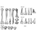
The present 3D Dataset contains the 3D models analyzed in the publication “Systematic and locomotor diversification of the Adapis group (Primates, Adapiformes) in the late Eocene of the Quercy (Southwest France), revealed by humeral remains”. In this paper, twenty humeral specimens from the old and new Quercy collections attributed to the fossil primates Adapis and Palaeolemur are described and analysed together. In this dataset only the scans of the fossils belonging to the collections of Université de Montpellier are provided.
In our paper (Marigó et al., 2019) we provide a qualitative and quantitative analysis of the different humeri, revealing that high variability is present within the “Adapis group” sample. Six different morphotypes are identified, confirming that what has often been called “Adapis parisiensis” is a mix of different species that present different locomotor adaptations.
Adapis sp. UM ROS 2-95 View specimen
|
M3#356Complete right humerus ROS 2-95 attributed to the Adapis group Type: "3D_surfaces"doi: 10.18563/m3.sf.356 state:published |
Download 3D surface file |
Adapis sp. UM ROS 2-536 View specimen
|
M3#357Proximal end of the right humerus ROS 2-536 attributed to the Adapis group Type: "3D_surfaces"doi: 10.18563/m3.sf.357 state:published |
Download 3D surface file |
Adapis sp. UM ROS 2-534 View specimen
|
M3#358Distal end of the left humerus ROS 2-534 attributed to the Adapis group Type: "3D_surfaces"doi: 10.18563/m3.sf.358 state:published |
Download 3D surface file |
Adapis sp. UM ROS 2-535 View specimen
|
M3#359Distal end of the left humerus ROS 2-535 attributed to the Adapis group Type: "3D_surfaces"doi: 10.18563/m3.sf.359 state:published |
Download 3D surface file |
Adapis sp. UM ROS 2-80 View specimen
|
M3#360Proximal end of the right humerus ROS 2-80 attributed to the Adapis group Type: "3D_surfaces"doi: 10.18563/m3.sf.360 state:published |
Download 3D surface file |
Adapis sp. UM ROS 2-79 View specimen
|
M3#361Distal end of the right humerus ROS 2-79 attributed to the Adapis group Type: "3D_surfaces"doi: 10.18563/m3.sf.361 state:published |
Download 3D surface file |
Adapis sp. UM ECA 1364 View specimen

|
M3#362Distal end of the left humerus ECA 1364 attributed to the Adapis group Type: "3D_surfaces"doi: 10.18563/m3.sf.362 state:published |
Download 3D surface file |
Adapis sp. UM ACQ-262 View specimen
|
M3#3733D model of ACQ 262. Humerus Type: "3D_surfaces"doi: 10.18563/m3.sf373 state:published |
Download 3D surface file |
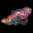
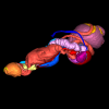
The present 3D Dataset contains the 3D models analyzed in: Hirose, A., Nakashima, T., Yamada, S., Uwabe, C., Kose, K., Takakuwa, T. 2012. Embryonic liver morphology and morphometry by magnetic resonance microscopic imaging. Anat Rec (Hoboken) 295, 51-59. doi: 10.1002/ar.21496
Homo sapiens KC-CS14LIV1387 View specimen
|
M3#64Human liver at Carnegie Stage (CS) 14 Type: "3D_surfaces"doi: 10.18563/m3.sf.64 state:published |
Download 3D surface file |
Homo sapiens KC-CS15LIV5074 View specimen
|
M3#65Human liver at Carnegie Stage (CS) 15 Type: "3D_surfaces"doi: 10.18563/m3.sf.65 state:published |
Download 3D surface file |
Homo sapiens KC-CS16LIV2578 View specimen
|
M3#66Human liver at Carnegie Stage (CS) 16 Type: "3D_surfaces"doi: 10.18563/m3.sf.66 state:published |
Download 3D surface file |
Homo sapiens KC-CS17LIV17832 View specimen
|
M3#67Human liver at Carnegie Stage (CS) 17 Type: "3D_surfaces"doi: 10.18563/m3.sf.67 state:published |
Download 3D surface file |
Homo sapiens KC-CS18LIV21124 View specimen
|
M3#68Human liver at Carnegie Stage (CS) 18 Type: "3D_surfaces"doi: 10.18563/m3.sf.68 state:published |
Download 3D surface file |
Homo sapiens KC-CS19LIV14353 View specimen

|
M3#69Human liver at Carnegie Stage (CS) 19 Type: "3D_surfaces"doi: 10.18563/m3.sf.69 state:published |
Download 3D surface file |
Homo sapiens KC-CS20LIV20701 View specimen
|
M3#70Human liver at Carnegie Stage (CS) 20 Type: "3D_surfaces"doi: 10.18563/m3.sf.70 state:published |
Download 3D surface file |
Homo sapiens KC-CS21LIV25858 View specimen
|
M3#71Human liver at Carnegie Stage (CS) 21 Type: "3D_surfaces"doi: 10.18563/m3.sf.71 state:published |
Download 3D surface file |
Homo sapiens KC-CS22LIV22226 View specimen
|
M3#72Human liver at Carnegie Stage (CS) 22 Type: "3D_surfaces"doi: 10.18563/m3.sf.72 state:published |
Download 3D surface file |
Homo sapiens KC-CS23LIV25704 View specimen
|
M3#73Human liver at Carnegie Stage (CS) 23 Type: "3D_surfaces"doi: 10.18563/m3.sf.73 state:published |
Download 3D surface file |
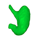
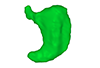
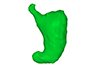
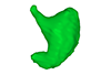
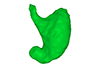
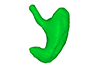
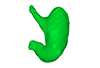
The present 3D Dataset contains the 3D models analyzed in: Kaigai N et al. Morphogenesis and three-dimensional movement of the stomach during the human embryonic period, Anat Rec (Hoboken). 2014 May;297(5):791-797. doi: 10.1002/ar.22833.
Homo sapiens KC-CS16STM27159 View specimen
|
M3#56computationally reconstructed stomach of the human embryo (M3#56_KC-CS16STM27159) at Carnegie Stage 16 (Crown Rump Length= 9.9mm). Type: "3D_surfaces"doi: 10.18563/m3.sf56 state:published |
Download 3D surface file |
Homo sapiens KC-CS17STM20383 View specimen
|
M3#57computationally reconstructed stomach of the human embryo (M3#57_KC-CS17STM20383) at Carnegie Stage 17 (Crown Rump Length= 12.3mm). Type: "3D_surfaces"doi: 10.18563/m3.sf57 state:published |
Download 3D surface file |
Homo sapiens KC-CS18STM21807 View specimen
|
M3#58computationally reconstructed stomach of the human embryo (M3#58_KC-CS18STM21807) at Carnegie Stage 18 (Crown Rump Length= 14.7mm). Type: "3D_surfaces"doi: 10.18563/m3.sf58 state:published |
Download 3D surface file |
Homo sapiens KC-CS19STM17998 View specimen
|
M3#59computationally reconstructed stomach of the human embryo (M3#59_KC-CS19STM17998) at Carnegie Stage 19 (Crown Rump Length was unmeasured ). Type: "3D_surfaces"doi: 10.18563/m3.sf59 state:published |
Download 3D surface file |
Homo sapiens KC-CS20STM20785 View specimen
|
M3#60computationally reconstructed stomach of the human embryo (M3#60_KC-CS20STM20785) at Carnegie Stage 20 (Crown Rump Length= 18.7 mm). Type: "3D_surfaces"doi: 10.18563/m3.sf60 state:published |
Download 3D surface file |
Homo sapiens KC-CS21STM24728 View specimen
|
M3#61computationally reconstructed stomach of the human embryo (M3#61_KC-CS21STM24728) at Carnegie Stage 21 (Crown Rump Length= 20.9 mm). Type: "3D_surfaces"doi: 10.18563/m3.sf61 state:published |
Download 3D surface file |
Homo sapiens KC-CS22STM26438 View specimen
|
M3#62computationally reconstructed stomach of the human embryo (M3#62_KC-CS22STM26438) at Carnegie Stage 22 (Crown Rump Length= 21.5 mm). Type: "3D_surfaces"doi: 10.18563/m3.sf62 state:published |
Download 3D surface file |
Homo sapiens KC-CS23STM20018 View specimen
|
M3#63computationally reconstructed stomach of the human embryo (M3#63_KC-CS23STM20018) at Carnegie Stage 23 (Crown Rump Length= 23.1 mm). Type: "3D_surfaces"doi: 10.18563/m3.sf63 state:published |
Download 3D surface file |
This project presents a µCT dataset and an associated 3D surface model of the holotype of Donrussellia magna (UM PAT 17; Primates, Adapiformes). UM PAT17 is the only known specimen for the species and consists of a well-preserved left lower jaw with p4-m3. It documents one of the oldest European primates, eventually dated near the Paleocene Eocene Thermal Maximum.
Donrussellia magna UM PAT 17 View specimen

|
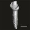
M3#173D surface file model of UM PAT 17 (type specimen of Donrussellia magna), which is a well preserved left lower jaw with p4-m3. The teeth (and roots) were manually segmented. Type: "3D_surfaces"doi: 10.18563/m3.sf17 state:published |
Download 3D surface file |
|
M3#18CT Scan Data of Donrussellia magna UM PAT 17. Voxel size (in µm): 36µm (isotropic voxels). Dimensions in x,y,z : 594 pixels, 294 pixels, 1038 pixels. Image type : 8-bit voxels. Image format : raw data format (no header). Type: "3D_CT"doi: 10.18563/m3.sf18 state:published |
Download CT data |
The 3D dataset presented in this article provides the 3D models of two Chelonioidea turtles dentaries from the Paleocene of France described in: Lapparent de Broin F. de, Marek H., Barrier P. & Gagnaison C. 2025. — Euclastidae n. fam. (Chelonioidea) et première mention d’Euclastes Cope, 1867 dans le Paléocène du bassin de Paris (France). Geodiversitas 47 (10): 409-464. https://doi.org/10.5252/geodiversitas2025v47a10.
Euclastes wielandi ULB-04A21-10 View specimen
|
M3#1791Euclastes wielandi Type: "3D_surfaces"doi: 10.18563/m3.sf.1791 state:published |
Download 3D surface file |
Euclastes wielandi MNHN.F.BPT52 View specimen
|
M3#1709Euclastes wielandi (cast) Type: "3D_surfaces"doi: 10.18563/m3.sf.1709 state:published |
Download 3D surface file |
Euclastes wielandi ULB-04A21-11 View specimen
|
M3#1792Euclastes montenati nov. sp. Type: "3D_surfaces"doi: 10.18563/m3.sf.1792 state:published |
Download 3D surface file |
Euclastes wielandi MNHN.F.BPT53 View specimen
|
M3#1711Euclastes montenati (cast) Type: "3D_surfaces"doi: 10.18563/m3.sf.1711 state:published |
Download 3D surface file |
The present 3D dataset contains the 3D models analyzed in the publication: Rosa, R. M., Salvador, R. B., & Cavallari, D. C. (2025). The disappearing act of the magician tree snail: anatomy, distribution, and phylogenetic relationships of Drymaeus magus (Gastropoda: Bulimulidae), a long-lost species hidden in plain sight. Zoological Journal of the Linnean Society.
Drymaeus magus CMRP 1049 View specimen
|
M3#1597Internal organs of Drymaeus magus Type: "3D_surfaces"doi: 10.18563/m3.sf.1597 state:published |
Download 3D surface file |

|
M3#1598External surface of Drymaeus magus Type: "3D_surfaces"doi: 10.18563/m3.sf.1598 state:published |
Download 3D surface file |
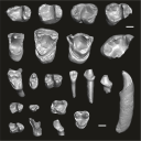
This contribution contains the three-dimensional digital models of the dental fossil material of anthropoid and strepsirrhine primates, discovered in Lower Oligocene detrital deposits outcropping in the Porto Rico and El Argoub areas, east of the Dakhla peninsula region (Atlantic Sahara; in the south of Morocco, near the northern border of Mauritania). These fossils were described, figured and discussed in the following publication: Marivaux et al. (2024), A new primate community from the earliest Oligocene of the Atlantic margin of Northwest Africa: Systematic, paleobiogeographic and paleoenvironmental implications. Journal of Human Evolution. https://doi.org/10.1016/j.jhevol.2024.103548
Catopithecus aff. browni DAK-Arg-087 View specimen

|
M3#1211Isolated right lower m3 (worn) Type: "3D_surfaces"doi: 10.18563/m3.sf.1211 state:published |
Download 3D surface file |
Catopithecus aff. browni DAK-Arg-088 View specimen
|
M3#1212Isolated right lower m2 (abraded/corroded) Type: "3D_surfaces"doi: 10.18563/m3.sf.1212 state:published |
Download 3D surface file |
Catopithecus aff. browni DAK-Arg-089 View specimen
|
M3#1213Isolated left lower m1 (worn) Type: "3D_surfaces"doi: 10.18563/m3.sf.1213 state:published |
Download 3D surface file |
Catopithecus aff. browni DAK-Pto-052 View specimen

|
M3#1214Isolated right lower m1 (pristine but lacking the mesiobuccal region) Type: "3D_surfaces"doi: 10.18563/m3.sf.1214 state:published |
Download 3D surface file |
Catopithecus aff. browni DAK-Arg-090 View specimen
|
M3#1215Isolated left upper P4 Type: "3D_surfaces"doi: 10.18563/m3.sf.1215 state:published |
Download 3D surface file |
Catopithecus aff. browni DAK-Arg-091 View specimen
|
M3#1216Isolated left upper M2 (worn and corroded) Type: "3D_surfaces"doi: 10.18563/m3.sf.1216 state:published |
Download 3D surface file |
Catopithecus aff. browni DAK-Pto-053 View specimen

|
M3#1217Isolated right upper M1 (lacking the buccal region) Type: "3D_surfaces"doi: 10.18563/m3.sf.1217 state:published |
Download 3D surface file |
Abuqatrania cf. basiodontos DAK-Arg-092 View specimen
|
M3#1218Isolated left lower c1 Type: "3D_surfaces"doi: 10.18563/m3.sf.1218 state:published |
Download 3D surface file |
?Propliopithecus sp. DAK-Pto-056 View specimen

|
M3#1219Isolated right lower m3 (fragment of talonid of a germ) Type: "3D_surfaces"doi: 10.18563/m3.sf.1219 state:published |
Download 3D surface file |
Abuqatrania cf. basiodontos DAK-Arg-093 View specimen

|
M3#1469Isolated right lower m1 Type: "3D_surfaces"doi: 10.18563/m3.sf.1469 state:published |
Download 3D surface file |
Abuqatrania cf. basiodontos DAK-Arg-094 View specimen
|
M3#1221Isolated left upper M1 or M2 (corroded, lacking the enamel cap [exposed dentine]) Type: "3D_surfaces"doi: 10.18563/m3.sf.1221 state:published |
Download 3D surface file |
Abuqatrania cf. basiodontos DAK-Arg-095 View specimen
|
M3#1222Isolated right lower i1 or i2 Type: "3D_surfaces"doi: 10.18563/m3.sf.1222 state:published |
Download 3D surface file |
Abuqatrania cf. basiodontos DAK-Arg-096 View specimen
|
M3#1223Isolated right lower p2 (worn apex) Type: "3D_surfaces"doi: 10.18563/m3.sf.1223 state:published |
Download 3D surface file |
Abuqatrania cf. basiodontos DAK-Arg-097 View specimen
|
M3#1224Isolated right lower p2 (worn apex and broken root) Type: "3D_surfaces"doi: 10.18563/m3.sf.1224 state:published |
Download 3D surface file |
Afrotarsius sp. DAK-Arg-098 View specimen

|
M3#1225Isolated left lower p3 Type: "3D_surfaces"doi: 10.18563/m3.sf.1225 state:published |
Download 3D surface file |
Afrotarsius sp. DAK-Pto-054 View specimen
|
M3#1226Isolated right lower m1 (abraded/corroded) Type: "3D_surfaces"doi: 10.18563/m3.sf.1226 state:published |
Download 3D surface file |
Orolemur mermozi DAK-Pto-055 View specimen
|
M3#1227Isolated right upper M1 or M2 (pristine, Holotype) Type: "3D_surfaces"doi: 10.18563/m3.sf.1227 state:published |
Download 3D surface file |
Wadilemur cf. elegans DAK-Arg-099 View specimen
|
M3#1228Isolated right lower m2 Type: "3D_surfaces"doi: 10.18563/m3.sf.1228 state:published |
Download 3D surface file |
cf. 'Anchomomys' milleri DAK-Arg-100 View specimen
|
M3#1229Isolated right lower c1 Type: "3D_surfaces"doi: 10.18563/m3.sf.1229 state:published |
Download 3D surface file |
Abuqatrania cf. basiodontos DAK-Arg-101 View specimen
|
M3#1396Isolated left upper M3 (abraded) Type: "3D_surfaces"doi: 10.18563/m3.sf.1396 state:published |
Download 3D surface file |
Orogalago saintexuperyi DAK-Arg-102 View specimen
|
M3#1397Isolated left lower m2 Type: "3D_surfaces"doi: 10.18563/m3.sf.1397 state:published |
Download 3D surface file |
Wadilemur cf. elegans DAK-Arg-103 View specimen
|
M3#1473Isolated right upper M1 or M2 (lacking the mesial and buccal regions) Type: "3D_surfaces"doi: 10.18563/m3.sf.1473 state:published |
Download 3D surface file |


