Explodable 3D Dog Skull for Veterinary Education
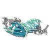
3D models of Ocnotherium skull
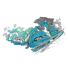
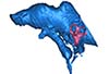
3D models of Kalakocetus, the earliest Cetacea
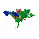
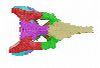
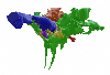
3D GM dataset of bird skeletal variation
Skeletal embryonic development in the catshark
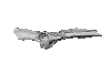
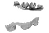
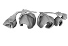
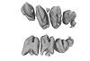
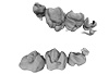
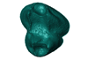
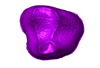
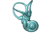
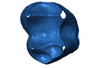
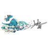
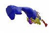
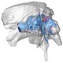
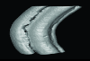
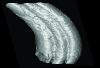
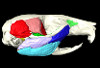
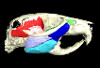
Bony connexions of the petrosal bone of extant hippos
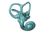
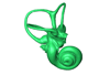
bony labyrinth (14) , inner ear (11) , geometric morphometrics (10) , CT-scan (10) , Eocene (10) , Micro-CT (9) , Miocene (8)
Lionel Hautier (24) , Maëva Judith Orliac (23) , Laurent Marivaux (18) , Renaud Lebrun (14) , Rodolphe Tabuce (14) , Pierre-Olivier Antoine (13) , Bastien Mennecart (13)
|
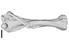
3D models related to the publication: Postcranial morphology of the extinct rodent Neoepiblema (Rodentia: Chinchilloidea): insights into the paleobiology of neoepiblemidsLeonardo Kerber
Published online: 20/10/2021 |
|
M3#719UFAC 3549, left humerus missing the proximal region. Type: "3D_surfaces"doi: 10.18563/m3.sf.719 state:published |
Download 3D surface file |
Neoepiblema acreensis UFAC 5076 View specimen
|
M3#720UFAC 5076, right humerus missing the proximal region. Type: "3D_surfaces"doi: 10.18563/m3.sf.720 state:published |
Download 3D surface file |
Neoepiblema acreensis UFAC 1939 View specimen
|
M3#721UFAC 1939, right ulna missing the olecranon epiphysis and the distal region. Type: "3D_surfaces"doi: 10.18563/m3.sf.721 state:published |
Download 3D surface file |
Neoepiblema acreensis UFAC 3697 View specimen
|
M3#722UFAC 3697, right innominate bone. Type: "3D_surfaces"doi: 10.18563/m3.sf.722 state:published |
Download 3D surface file |
Neoepiblema acreensis UFAC 2574 View specimen
|
M3#723UFAC 2574, proximal region of a left femur. Type: "3D_surfaces"doi: 10.18563/m3.sf.723 state:published |
Download 3D surface file |
Neoepiblema acreensis UFAC 2937 View specimen
|
M3#724UFAC 2937, right femur with damaged proximal region. Type: "3D_surfaces"doi: 10.18563/m3.sf.724 state:published |
Download 3D surface file |
Neoepiblema acreensis UFAC 2210 View specimen
|
M3#725UFAC 2210, distal region of a right femur. Type: "3D_surfaces"doi: 10.18563/m3.sf.725 state:published |
Download 3D surface file |
Neoepiblema acreensis UFAC 1887 View specimen
|
M3#726UFAC 1887, right tibia Type: "3D_surfaces"doi: 10.18563/m3.sf.726 state:published |
Download 3D surface file |
Neoepiblema acreensis UFAC 1840 View specimen
|
M3#727UFAC 1840, left astragalus. Type: "3D_surfaces"doi: 10.18563/m3.sf.727 state:published |
Download 3D surface file |
Neoepiblema acreensis UFAC 2549 View specimen
|
M3#728UFAC 2549, right astragalus. Type: "3D_surfaces"doi: 10.18563/m3.sf.728 state:published |
Download 3D surface file |
Neoepiblema acreensis UFAC 3672 View specimen
|
M3#729UFAC 3672, right navicular. Type: "3D_surfaces"doi: 10.18563/m3.sf.729 state:published |
Download 3D surface file |
Neoepiblema acreensis UFAC 2116 View specimen
|
M3#730UFAC 2116, left metatarsal III. Type: "3D_surfaces"doi: 10.18563/m3.sf.730 state:published |
Download 3D surface file |
Neoepiblema horridula UFAC 3260 View specimen
|
M3#731UFAC 3260, fragmented left innominate. Type: "3D_surfaces"doi: 10.18563/m3.sf.731 state:published |
Download 3D surface file |
Neoepiblema horridula UFAC 2620 View specimen
|
M3#732UFAC 2620, distal region of a right femur. Type: "3D_surfaces"doi: 10.18563/m3.sf.732 state:published |
Download 3D surface file |
Neoepiblema horridula UFAC 2737 View specimen
|
M3#733UFAC 2737, proximal region of right femur. Type: "3D_surfaces"doi: 10.18563/m3.sf.733 state:published |
Download 3D surface file |
Neoepiblema horridula UFAC 3202 View specimen
|
M3#734UFAC 3202, right tibia, missing the proximalmost and distal portions. Type: "3D_surfaces"doi: 10.18563/m3.sf.734 state:published |
Download 3D surface file |
Neoepiblema horridula UFAC 3212 View specimen
|
M3#735UFAC 3212, left astragalus. Type: "3D_surfaces"doi: 10.18563/m3.sf.735 state:published |
Download 3D surface file |
The present 3D Dataset contains the 3D model of the brain endocast of Neoepiblema acreensis analyzed in “Small within the largest: Brain size and anatomy of the extinct Neoepiblema acreensis, a giant rodent from the Neotropics”. The 3D model was generated using CT-Scanning and techniques of virtual reconstruction.
Neoepiblema acreensis UFAC 4515 View specimen
|
M3#502Brain endocast of Neoepiblema acreensis Type: "3D_surfaces"doi: 10.18563/m3.sf.502 state:published |
Download 3D surface file |
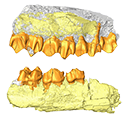
The present 3D Dataset contains the 3D models analyzed in ”Morphological features of tooth development and replacement in the rabbit Oryctolagus cuniculus”, Archives of Oral Biology, https://doi.org/10.1016/j.archoralbio.2019.104576
Oryctogalus cuniculus E14 View specimen
|
M3#390Right cheek teeth, Left and right incisors at 14 dpf Type: "3D_surfaces"doi: 10.18563/m3.sf.390 state:published |
Download 3D surface file |
Oryctogalus cuniculus E16 View specimen
|
M3#391Left cheek teeth, Left and right incisors at 16 dpf Type: "3D_surfaces"doi: 10.18563/m3.sf.391 state:published |
Download 3D surface file |
Oryctogalus cuniculus E18 View specimen
|
M3#392Left cheek teeth and incisors at 18 dpf Type: "3D_surfaces"doi: 10.18563/m3.sf.392 state:published |
Download 3D surface file |
Oryctogalus cuniculus E20 View specimen
|
M3#393Left cheek teeth and incisors at 20 dpf Type: "3D_surfaces"doi: 10.18563/m3.sf.393 state:published |
Download 3D surface file |
Oryctogalus cuniculus E22 View specimen
|
M3#394Left lower cheek teeth and incisors, right upper cheek teeth and incisors at 22 dpf Type: "3D_surfaces"doi: 10.18563/m3.sf.394 state:published |
Download 3D surface file |
Oryctogalus cuniculus E24 View specimen
|
M3#395Left cheek teeth and incisors at 24 dpf Type: "3D_surfaces"doi: 10.18563/m3.sf.395 state:published |
Download 3D surface file |
Oryctogalus cuniculus E28 View specimen
|
M3#396Right cheek teeth and incisors at 28 dpf Type: "3D_surfaces"doi: 10.18563/m3.sf.396 state:published |
Download 3D surface file |
Oryctogalus cuniculus E26 View specimen
|
M3#397Right cheek teeth and incisors at 26 dpf Type: "3D_surfaces"doi: 10.18563/m3.sf.397 state:published |
Download 3D surface file |
The present 3D Dataset contains the 3D models of the enamel-dentine junctions of upper third molars and of the bony labyrinths of the extant cercopithecoid specimens analyzed in the following publication: Beaudet, A., Dumoncel, J., Thackeray, J.F., Bruxelles, L., Duployer, B., Tenailleau, C., Bam, L., Hoffman, J., de Beer, F., Braga, J.: Upper third molar internal structural organization and semicircular canal morphology in Plio-Pleistocene South African cercopithecoids. Journal of Human Evolution 95, 104-120. https://doi.org/10.1016/j.jhevol.2016.04.004
Cercocebus atys 81.007-M-0041 View specimen
|
M3#4453D model of the enamel-dentine junction of the right upper third molar. Type: "3D_surfaces"doi: 10.18563/m3.sf.445 state:published |
Download 3D surface file |
Cercocebus torquatus 73.018-M-0359 View specimen
|
M3#4463D model of the enamel-dentine junction of the right upper third molar. Type: "3D_surfaces"doi: 10.18563/m3.sf.446 state:published |
Download 3D surface file |

|
M3#4963D model of the left bony labyrinth. Type: "3D_surfaces"doi: 10.18563/m3.sf.496 state:published |
Download 3D surface file |
Mandrillus leucophaeus 73.029-M-0106 View specimen
|
M3#4473D model of the enamel-dentine junction of the right upper third molar. Type: "3D_surfaces"doi: 10.18563/m3.sf.447 state:published |
Download 3D surface file |

|
M3#4703D model of the right bony labyrinth. Type: "3D_surfaces"doi: 10.18563/m3.sf.470 state:published |
Download 3D surface file |
Lophocebus albigena 73.029-M-0109 View specimen
|
M3#4483D model of the enamel-dentine junction of the right upper third molar. Type: "3D_surfaces"doi: 10.18563/m3.sf.448 state:published |
Download 3D surface file |
|
M3#4713D model of the right bony labyrinth. Type: "3D_surfaces"doi: 10.18563/m3.sf.471 state:published |
Download 3D surface file |
Piliocolobus foai 91.060-M-0071 View specimen
|
M3#4493D model of the enamel-dentine junction of the right upper third molar. Type: "3D_surfaces"doi: 10.18563/m3.sf.449 state:published |
Download 3D surface file |

|
M3#4723D model of the right bony labyrinth. Type: "3D_surfaces"doi: 10.18563/m3.sf.472 state:published |
Download 3D surface file |
Colobus guereza 1215 View specimen
|
M3#4503D model of the enamel-dentine junction of the right upper third molar. Type: "3D_surfaces"doi: 10.18563/m3.sf.450 state:published |
Download 3D surface file |

|
M3#4733D model of the right bony labyrinth. Type: "3D_surfaces"doi: 10.18563/m3.sf.473 state:published |
Download 3D surface file |
Colobus guereza 2800 View specimen
|
M3#4513D model of the enamel-dentine junction of the right upper third molar. Type: "3D_surfaces"doi: 10.18563/m3.sf.451 state:published |
Download 3D surface file |

|
M3#4743D model of the right bony labyrinth. Type: "3D_surfaces"doi: 10.18563/m3.sf.474 state:published |
Download 3D surface file |
Papio cynocephalus kindae 3503 View specimen
|
M3#4523D model of the enamel-dentine junction of the right upper third molar. Type: "3D_surfaces"doi: 10.18563/m3.sf.452 state:published |
Download 3D surface file |

|
M3#4753D model of the right bony labyrinth. Type: "3D_surfaces"doi: 10.18563/m3.sf.475 state:published |
Download 3D surface file |
Erythrocebus patas 8452 View specimen
|
M3#4533D model of the enamel-dentine junction of the right upper third molar. Type: "3D_surfaces"doi: 10.18563/m3.sf.453 state:published |
Download 3D surface file |

|
M3#4763D model of the right bony labyrinth. Type: "3D_surfaces"doi: 10.18563/m3.sf.476 state:published |
Download 3D surface file |
Papio cynocephalus kindae 17979 View specimen
|
M3#4543D model of the enamel-dentine junction of the right upper third molar. Type: "3D_surfaces"doi: 10.18563/m3.sf.454 state:published |
Download 3D surface file |

|
M3#4773D model of the right bony labyrinth. Type: "3D_surfaces"doi: 10.18563/m3.sf.477 state:published |
Download 3D surface file |
Colobus angolensis 25456 View specimen
|
M3#4553D model of the enamel-dentine junction of the right upper third molar. Type: "3D_surfaces"doi: 10.18563/m3.sf.455 state:published |
Download 3D surface file |

|
M3#4783D model of the right bony labyrinth. Type: "3D_surfaces"doi: 10.18563/m3.sf.478 state:published |
Download 3D surface file |
Chlorocebus pygerythrus 37477 View specimen
|
M3#4563D model of the enamel-dentine junction of the right upper third molar. Type: "3D_surfaces"doi: 10.18563/m3.sf.456 state:published |
Download 3D surface file |
|
M3#4813D model of the right bony labyrinth. Type: "3D_surfaces"doi: 10.18563/m3.sf.481 state:published |
Download 3D surface file |
Chlorocebus pygerythrus 37478 View specimen
|
M3#4573D model of the enamel-dentine junction of the right upper third molar. Type: "3D_surfaces"doi: 10.18563/m3.sf.457 state:published |
Download 3D surface file |

|
M3#4823D model of the right bony labyrinth. Type: "3D_surfaces"doi: 10.18563/m3.sf.482 state:published |
Download 3D surface file |
Lophocebus albigena 37572 View specimen
|
M3#4583D model of the enamel-dentine junction of the right upper third molar. Type: "3D_surfaces"doi: 10.18563/m3.sf.458 state:published |
Download 3D surface file |

|
M3#4833D model of the right bony labyrinth. Type: "3D_surfaces"doi: 10.18563/m3.sf.483 state:published |
Download 3D surface file |
Lophocebus albigena 37579 View specimen
|
M3#4593D model of the enamel-dentine junction of the right upper third molar. Type: "3D_surfaces"doi: 10.18563/m3.sf.459 state:published |
Download 3D surface file |
Erythrocebus patas OST.2002-26 View specimen
|
M3#4603D model of the enamel-dentine junction of the right upper third molar. Type: "3D_surfaces"doi: 10.18563/m3.sf.460 state:published |
Download 3D surface file |
|
M3#4843D model of the right bony labyrinth. Type: "3D_surfaces"doi: 10.18563/m3.sf.484 state:published |
Download 3D surface file |
Mandrillus sphinx OST.AC.488 View specimen
|
M3#4613D model of the enamel-dentine junction of the right upper third molar. Type: "3D_surfaces"doi: 10.18563/m3.sf.461 state:published |
Download 3D surface file |
|
M3#4853D model of the left bony labyrinth. Type: "3D_surfaces"doi: 10.18563/m3.sf.485 state:published |
Download 3D surface file |
Macaca mulatta OST.AC.492 View specimen
|
M3#4623D model of the enamel-dentine junction of the right upper third molar. Type: "3D_surfaces"doi: 10.18563/m3.sf.462 state:published |
Download 3D surface file |
|
M3#4863D model of the right bony labyrinth. Type: "3D_surfaces"doi: 10.18563/m3.sf.486 state:published |
Download 3D surface file |
Chlorocebus aethiops OST.AC.523 View specimen
|
M3#4633D model of the enamel-dentine junction of the right upper third molar. Type: "3D_surfaces"doi: 10.18563/m3.sf.463 state:published |
Download 3D surface file |

|
M3#4913D model of the right bony labyrinth. Type: "3D_surfaces"doi: 10.18563/m3.sf.491 state:published |
Download 3D surface file |
Cercopithecus cephus OST.AC.533 View specimen
|
M3#4643D model of the enamel-dentine junction of the right upper third molar. Type: "3D_surfaces"doi: 10.18563/m3.sf.464 state:published |
Download 3D surface file |
|
M3#4933D model of the right bony labyrinth. Type: "3D_surfaces"doi: 10.18563/m3.sf.493 state:published |
Download 3D surface file |
Chlorocebus aethiops OST.AC.540 View specimen
|
M3#4653D model of the enamel-dentine junction of the right upper third molar. Type: "3D_surfaces"doi: 10.18563/m3.sf.465 state:published |
Download 3D surface file |

|
M3#4943D model of the right bony labyrinth. Type: "3D_surfaces"doi: 10.18563/m3.sf.494 state:published |
Download 3D surface file |
Mandrillus sphinx OST.AC.543 View specimen
|
M3#4663D model of the enamel-dentine junction of the right upper third molar. Type: "3D_surfaces"doi: 10.18563/m3.sf.466 state:published |
Download 3D surface file |
|
M3#4953D model of the right bony labyrinth. Type: "3D_surfaces"doi: 10.18563/m3.sf.495 state:published |
Download 3D surface file |
Cercocebus torquatus 73.018-M-389 View specimen

|
M3#4683D model of the right bony labyrinth. Type: "3D_surfaces"doi: 10.18563/m3.sf.468 state:published |
Download 3D surface file |
Mandrillus leucophaeus 73.029-M-0105 View specimen
|
M3#4693D model of the right bony labyrinth. Type: "3D_surfaces"doi: 10.18563/m3.sf.469 state:published |
Download 3D surface file |
Mandrillus leucophaeus 28425 View specimen

|
M3#4793D model of the right bony labyrinth. Type: "3D_surfaces"doi: 10.18563/m3.sf.479 state:published |
Download 3D surface file |
Cercocebus atys 28998 View specimen
|
M3#4803D model of the right bony labyrinth. Type: "3D_surfaces"doi: 10.18563/m3.sf.480 state:published |
Download 3D surface file |
Macaca sylvanus OST.AC.493 View specimen

|
M3#4873D model of the right bony labyrinth. Type: "3D_surfaces"doi: 10.18563/m3.sf.487 state:published |
Download 3D surface file |
Chlorocebus aethiops OST.AC.508 View specimen

|
M3#4883D model of the left bony labyrinth. Type: "3D_surfaces"doi: 10.18563/m3.sf.488 state:published |
Download 3D surface file |
Cercopithecus cephus OST.AC.515 View specimen

|
M3#4893D model of the right bony labyrinth. Type: "3D_surfaces"doi: 10.18563/m3.sf.489 state:published |
Download 3D surface file |
Colobus guereza OST.AC.519 View specimen

|
M3#4903D model of the right bony labyrinth. Type: "3D_surfaces"doi: 10.18563/m3.sf.490 state:published |
Download 3D surface file |
Macaca sp. OST.AC.532 View specimen

|
M3#4923D model of the left bony labyrinth. Type: "3D_surfaces"doi: 10.18563/m3.sf.492 state:published |
Download 3D surface file |
The present 3D Dataset contains the 3D model analyzed in the article : Dubied et al. (2021), Endocranium and ecology of Eurotherium theriodis, a European hyaenodont mammal from the Lutetian. Acta Palaeontologica Polonica 2021, https://doi.org/10.4202/app.00771.2020
Eurotherium theriodis NMB.Em12 View specimen
|
M3#381NMB.Em12 unprepared specimen Type: "3D_surfaces"doi: 10.18563/m3.sf.381 state:published |
Download 3D surface file |
|
M3#382NMB.Em12 cranium Type: "3D_surfaces"doi: 10.18563/m3.sf.382 state:published |
Download 3D surface file |
|
M3#383NMB.Em12 endocast Type: "3D_surfaces"doi: 10.18563/m3.sf.383 state:published |
Download 3D surface file |
This contribution contains the 3D models described and figured in: The Neogene record of northern South American native ungulates. Smithsonian Contributions to Paleobiology. Doi: 10.5479/si.1943-6688.101
Hilarcotherium miyou IGMp 881327 View specimen
|
M3#318Right upper M2 Type: "3D_surfaces"doi: 10.18563/m3.sf.318 state:published |
Download 3D surface file |
Hilarcotherium miyou MUN-STRI 34216 View specimen
|
M3#319Right upper P4 Type: "3D_surfaces"doi: 10.18563/m3.sf.319 state:published |
Download 3D surface file |
|
M3#320Right upper M2 Type: "3D_surfaces"doi: 10.18563/m3.sf.320 state:published |
Download 3D surface file |
Falcontoxodon aguilerai AMU-CURS 585 View specimen
|
M3#321Maxilla with left M3-P2 and right I2 Type: "3D_surfaces"doi: 10.18563/m3.sf.321 state:published |
Download 3D surface file |
The present 3D Dataset contains the 3D models analyzed in: Toyoda S et al., 2015, Morphogenesis of the inner ear at different stages of normal human development. The Anatomical Record. doi : 10.1002/ar.23268
Homo sapiens KC-CS17IER29248 View specimen

|
M3#36Computationally reconstructed membranous labyrinth of a human embryo (KC-CS17IER29248) at Carnegie Stage 17 (Crown Rump Length= 7mm). Type: "3D_surfaces"doi: 10.18563/m3.sf36 state:published |
Download 3D surface file |
Homo sapiens KC-CS18IER17746 View specimen

|
M3#37Computationally reconstructed membranous labyrinth of a human embryo (KC-CS18IER17746) at Carnegie Stage 18 (Crown Rump Length= 12mm). Type: "3D_surfaces"doi: 10.18563/m3.sf37 state:published |
Download 3D surface file |
Homo sapiens KC-CS19IER16127 View specimen

|
M3#38Computationally reconstructed membranous labyrinth of a human embryo (KC-CS19IER16127) at Carnegie Stage 19 (Crown Rump Length= 13mm). Type: "3D_surfaces"doi: 10.18563/m3.sf38 state:published |
Download 3D surface file |
Homo sapiens KC-CS20IER20268 View specimen

|
M3#39Computationally reconstructed membranous labyrinth of a human embryo (KC-CS20IER20268) at Carnegie Stage 20 (Crown Rump Length= 13.7mm). Type: "3D_surfaces"doi: 10.18563/m3.sf39 state:published |
Download 3D surface file |
Homo sapiens KC-CS21IER28066 View specimen

|
M3#40Computationally reconstructed membranous labyrinth of a human embryo (KC-CS21IER28066) at Carnegie Stage 21 (Crown Rump Length= 16.7mm). Type: "3D_surfaces"doi: 10.18563/m3.sf40 state:published |
Download 3D surface file |
Homo sapiens KC-CS22IER35233 View specimen

|
M3#41Computationally reconstructed membranous labyrinth of a human embryo (KC-CS22IER35233) at Carnegie Stage 22 (Crown Rump Length= 22mm). Type: "3D_surfaces"doi: 10.18563/m3.sf41 state:published |
Download 3D surface file |
Homo sapiens KC-CS23IER15919 View specimen

|
M3#42Computationally reconstructed membranous labyrinth of a human embryo (KC-CS23IER15919) at Carnegie Stage 23 (Crown Rump Length= 32.3mm). Type: "3D_surfaces"doi: 10.18563/m3.sf42 state:published |
Download 3D surface file |
Homo sapiens KC-FIER52730 View specimen

|
M3#43Computationally reconstructed human membranous labyrinth in post embryonic phase (KC-FIER52730). Crown Rump Length: 43.5mm. Type: "3D_surfaces"doi: 10.18563/m3.sf43 state:published |
Download 3D surface file |
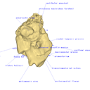
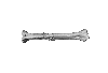
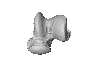
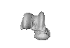
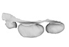
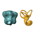
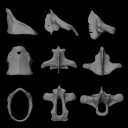
This project presents the 3D models of two isolated petrosals from the Oligocene locality of Pech de Fraysse (Quercy, France) here attributed to the genus Prodremotherium Filhol, 1877. Our aim is to describe the petrosal morphology of this Oligocene “early ruminant” as only few data are available in the literature for Oligocene taxa.
Prodremotherium sp. UM PFY 4053 View specimen

|
M3#7Labelled 3D model of right isolated petrosal of Prodremotherium sp. from Pech de Fraysse (Quercy, MP 28) Type: "3D_surfaces"doi: 10.18563/m3.sf7 state:published |
Download 3D surface file |
Prodremotherium sp. UM PFY 4054 View specimen

|
M3#8Labelled 3D model of right isolated petrosal of Prodremotherium sp. from Pech de Fraysse (Quercy, MP 28) Type: "3D_surfaces"doi: 10.18563/m3.sf8 state:published |
Download 3D surface file |

This contribution contains 3D models of the cranial endoskeleton of three specimens of the Permian ‘acanthodian’ stem-group chondrichthyan (cartilaginous fish) Acanthodes confusus, obtained using computed tomography. These datasets were described and analyzed in Dearden et al. (2024) “3D models related to the publication: The pharynx of the iconic stem-group chondrichthyan Acanthodes Agassiz, 1833 revisited with micro computed tomography.” Zoological Journal of the Linnean Society
Acanthodes confusus MNHN-F-SAA20 View specimen
|
M3#14703D surfaces representing the three-dimensionally fossilised head of Acanthodes confusus Type: "3D_surfaces"doi: 10.18563/m3.sf.1470 state:published |
Download 3D surface file |
Acanthodes confusus MNHN-F-SAA21 View specimen
|
M3#14713D surfaces representing the three-dimensionally fossilised head of Acanthodes confusus Type: "3D_surfaces"doi: 10.18563/m3.sf.1471 state:published |
Download 3D surface file |
Acanthodes confusus MNHN-F-SAA24 View specimen
|
M3#14723D surfaces representing the three-dimensionally fossilised head of Acanthodes confusus Type: "3D_surfaces"doi: 10.18563/m3.sf.1472 state:published |
Download 3D surface file |
The present 3D Dataset contains the 3D models analyzed in Brualla et al., 2024: Comparative anatomy of the vocal apparatus in bats and implication for the diversity of laryngeal echolocation. Zoological Journal of the Linnean Society, vol. zlad180. (https://doi.org/10.1093/zoolinnean/zlad180). Bat larynges are understudied in the previous anatomical studies. The description and comparison of the different morphological traits might provide important proxies to investigate the evolutionary origin of laryngeal echolocation in bats.
Eonycteris spelaea VN18-026 View specimen

|
M3#1305Laryngeal cartilages and muscles of the cave nectar bat Type: "3D_surfaces"doi: 10.18563/m3.sf.1305 state:published |
Download 3D surface file |
Macroglossus sobrinus VN15-017 View specimen
|
M3#1306Laryngeal anatomy of Macroglossus sobrinus Type: "3D_surfaces"doi: 10.18563/m3.sf.1306 state:published |
Download 3D surface file |
Aselliscus dongbacana VTTu15-013 View specimen
|
M3#1307Laryngeal anatomy of Aselliscus dongbacana Type: "3D_surfaces"doi: 10.18563/m3.sf.1307 state:published |
Download 3D surface file |
Coelops frithii VN19-196 View specimen
|
M3#1308Laryngeal anatomy of Coelops frithii Type: "3D_surfaces"doi: 10.18563/m3.sf.1308 state:published |
Download 3D surface file |
Hipposideros larvatus VN18-209 View specimen
|
M3#1309Laryngeal anatomy of Hipposideros larvatus Type: "3D_surfaces"doi: 10.18563/m3.sf.1309 state:published |
Download 3D surface file |
Rhinolophus cornutus JP21-025 View specimen

|
M3#14763D surfaces of Rhinolophus cornutus Type: "3D_surfaces"doi: 10.18563/m3.sf.1476 state:published |
Download 3D surface file |
Rhinolophus macrotis VN11-089 View specimen
|
M3#1477Laryngeal cartilages and muscles of Rhinolophus macrotis Type: "3D_surfaces"doi: 10.18563/m3.sf.1477 state:published |
Download 3D surface file |
Lyroderma lyra VN17-535 View specimen
|
M3#1312Laryngeal anatomy of Lyroderma lyra Type: "3D_surfaces"doi: 10.18563/m3.sf.1312 state:published |
Download 3D surface file |
Saccolaimus mixtus A3257 View specimen
|
M3#1478Laryngeal components of Saccolaimus mixtus Type: "3D_surfaces"doi: 10.18563/m3.sf.1478 state:published |
Download 3D surface file |
Taphozous melanopogon VN17-0252 View specimen
|
M3#1479Laryngeal cartilages and muscles of Taphozous melanopogon Type: "3D_surfaces"doi: 10.18563/m3.sf.1479 state:published |
Download 3D surface file |
Artibeus jamaicensis AJ001 View specimen
|
M3#1316Laryngeal anatomy of Artibeus jamaicensis Type: "3D_surfaces"doi: 10.18563/m3.sf.1316 state:published |
Download 3D surface file |
Kerivoula hardwickii VN11-0043 View specimen
|
M3#1317Laryngeal anatomy of Kerivoula hardwickii Type: "3D_surfaces"doi: 10.18563/m3.sf.1317 state:published |
Download 3D surface file |
Myotis ater VN19-016 View specimen
|
M3#1318Laryngeal anatomy of Myotis ater Type: "3D_surfaces"doi: 10.18563/m3.sf.1318 state:published |
Download 3D surface file |
Myotis siligorensis VTTu14-018 View specimen
|
M3#1319Laryngeal anatomy of Myotis siligorensis Type: "3D_surfaces"doi: 10.18563/m3.sf.1319 state:published |
Download 3D surface file |
Suncus murinus KATS_835A View specimen
|
M3#1395Laryngeal anatomy of Suncus murinus Type: "3D_surfaces"doi: 10.18563/m3.sf.1395 state:published |
Download 3D surface file |
The present 3D Dataset contains the 3D model analyzed in the following publication: occurrence of the ground sloth Nothrotheriops (Xenarthra, Folivora) in the Late Pleistocene of Uruguay: New information on its dietary and habitat preferences based on stable isotope analysis. Journal of Mammalian Evolution. https://doi.org/10.1007/s10914-023-09660-w
Nothrotheriops sp. CAV 1466 View specimen
|
M3#1129Left humerus Type: "3D_surfaces"doi: 10.18563/m3.sf.1129 state:published |
Download 3D surface file |
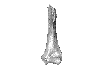
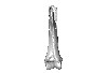
The present 3D Dataset contains 3D models of the holotypes described in Aiglstorfer et al. (2023a). Miocene Moschidae (Mammalia, Ruminantia) from the Linxia Basin (China) connect Europe and Asia and show early evolutionary diversity of a today monogeneric family. Palaeogeography, Palaeoclimatology, Palaeoecology.
Micromeryx? caoi CUGB GV 87045 View specimen

|
M3#11123D models of the holotype of “Micromeryx” caoi (CUGB GV87045) including the models of the teeth, the mandibule, and the sediment. Type: "3D_surfaces"doi: 10.18563/m3.sf.1112 state:published |
Download 3D surface file |
Hispanomeryx linxiaensis IVPP V28596 View specimen

|
M3#11133D models of the holotype of Hispanomeryx linxiaensis (IVPP V28596) including the models of the teeth, the mandibule, and the sediment. Type: "3D_surfaces"doi: 10.18563/m3.sf.1113 state:published |
Download 3D surface file |
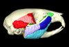
The present 3D Dataset contains the 3D models analyzed in Pochat-Cottilloux Y., Rinder N., Perrichon G., Adrien J., Amiot R., Hua S. & Martin J. E. (2023). The neuroanatomy and pneumaticity of Hamadasuchus from the Cretaceous of Morocco and its significance for the paleoecology of Peirosauridae and other altirostral crocodylomorphs. Journal of Anatomy, https://doi.org/10.1111/joa.13887
Hamadasuchus sp. UCBL-FSL 532408 View specimen
|
M3#10943D volume reconstruction of the braincase osteology Type: "3D_surfaces"doi: 10.18563/m3.sf.1094 state:published |
Download 3D surface file |
|
M3#10963D volume reconstruction of the endocast Type: "3D_surfaces"doi: 10.18563/m3.sf.1096 state:published |
Download 3D surface file |
|
M3#10973D volume reconstruction of the labyrinths Type: "3D_surfaces"doi: 10.18563/m3.sf.1097 state:published |
Download 3D surface file |
|
M3#10983D volume reconstruction of the pneumatic cavities Type: "3D_surfaces"doi: 10.18563/m3.sf.1098 state:published |
Download 3D surface file |
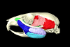
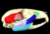
This contribution contains the 3D models described and figured in the following publication: Paulina-Carabajal A and Calvo JO 2021. Re-description of the braincase of the rebbachisaurid sauropod Limaysaurus tessonei and novel endocranial information based on CT scans. Anais da Academia Brasileira de Ciências 93(Suppl. 2): e20200762 https://doi.org/10.1590/0001-3765202120200762
Limaysaurus tessonei MUCPv-205 View specimen
|
M3#700Renderings of the virtually isolate braincase, brain, and right inner ear. Type: "3D_surfaces"doi: 10.18563/m3.sf.700 state:published |
Download 3D surface file |
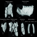
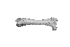
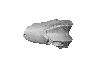
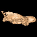
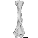
This contribution contains 3D models of extinct rodents Dinomyidae from Miocene and Quaternary of Brazil. The Miocene specimens that were digitalized include the holotypes of Potamarchus adamiae, Pseudopotamarchus villanuevai, and Ferigolomys pacarana collected in the Solimões Formation (Upper Miocene), northern Brazil. The Quaternary specimens are the holotype and paratype of Niedemys piauiensis, found in Upper Pleistocene deposits from northeast Brazil.
Potamarchus adamiae UFAC-CS 011 View specimen
|
M3#410UFAC-CS 011 – holotype, palatal region of the skull with cheek teeth Type: "3D_surfaces"doi: 10.18563/m3.sf.410 state:published |
Download 3D surface file |
Potamarchus adamiae UFAC-CS 043 View specimen
|
M3#411UFAC-CS 043, left dentary with cheek teeth Type: "3D_surfaces"doi: 10.18563/m3.sf.411 state:published |
Download 3D surface file |
Pseudopotamarchus villanuevai UFAC 4762 View specimen
|
M3#412UFAC 4762 – holotype, incomplete right maxilla with cheek teeth Type: "3D_surfaces"doi: 10.18563/m3.sf.412 state:published |
Download 3D surface file |
Ferigolomys pacarana UFAC 6460 View specimen
|
M3#413UFAC 6460 – holotype, palatal region of the skull with cheek teeth Type: "3D_surfaces"doi: 10.18563/m3.sf.413 state:published |
Download 3D surface file |
Drytomomys sp. UFAC 2742 View specimen

|
M3#414UFAC 2742, right dentary with cheek teeth Type: "3D_surfaces"doi: 10.18563/m3.sf.414 state:published |
Download 3D surface file |
Niedemys piauiensis FUMDHAM 113-146365-2 View specimen
|
M3#418FUMDHAM 113-146365-2 - holotype, upper right tooth Type: "3D_surfaces"doi: 10.18563/m3.sf.418 state:published |
Download 3D surface file |
Niedemys piauiensis FUMDHAM 113-145304-2 View specimen
|
M3#419FUMDHAM 113-145304-2 - paratype, left lower molar Type: "3D_surfaces"doi: 10.18563/m3.sf.419 state:published |
Download 3D surface file |
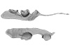
This contribution contains the 3D model of the fossil talus of a small-bodied anthropoid primate (Platyrrhini, Cebidae, Cebinae) discovered from lower Miocene deposits of Peruvian Amazonia (MD-61 locality, Upper Madre de Dios Basin). This fossil was described and figured in the following publication: Marivaux et al. (2012), A platyrrhine talus from the early Miocene of Peru (Amazonian Madre de Dios Sub-Andean Zone). Journal of Human Evolution. http://dx.doi.org/10.1016/j.jhevol.2012.07.005
Cebinae indet. sp. MUSM-2024 View specimen
|
M3#380Right talus 3D surface of a Miocene Cebinae indet. primate Type: "3D_surfaces"doi: 10.18563/m3.sf.380 state:published |
Download 3D surface file |
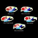
The present 3D Dataset contains the 3D models analyzed in the article entitled "One skull to rule them all? Descriptive and comparative anatomy of the masticatory apparatus in five mice species based on traditional and digital dissections" (Ginot et al. 2018, Journal of Morphology, https://doi.org/10.1002/jmor.20845).
Mus cervicolor R7314 View specimen
|
M3#343.ply surfaces of the skull and masticatory muscles of Mus cervicolor. Created with MorphoDig, .pos and .ntw files also included. Scans were obtained thanks to the Institut des Sciences de l'Evolution de Montpellier MRI platform. Type: "3D_surfaces"doi: 10.18563/m3.sf.343 state:published |
Download 3D surface file |
Mus caroli R7264 View specimen
|
M3#344.ply surfaces of the skull and masticatory muscles of Mus caroli. Created with MorphoDig, .pos and .ntw files also included. Scans were obtained thanks to the Institut des Sciences de l'Evolution de Montpellier MRI platform. Type: "3D_surfaces"doi: 10.18563/m3.sf.344 state:published |
Download 3D surface file |
Mus fragilicauda R7260 View specimen
|
M3#345.ply surfaces of the skull and masticatory muscles of Mus fragilicauda. Created with MorphoDig, .pos and .ntw files also included. Scans were obtained thanks to the Institut des Sciences de l'Evolution de Montpellier MRI platform. Type: "3D_surfaces"doi: 10.18563/m3.sf.345 state:published |
Download 3D surface file |
Mus pahari R7226 View specimen
|
M3#346.ply surfaces of the skull and masticatory muscles of Mus pahari. Created with MorphoDig, .pos and .ntw files also included. Scans were obtained thanks to the Institut des Sciences de l'Evolution de Montpellier MRI platform. Type: "3D_surfaces"doi: 10.18563/m3.sf.346 state:published |
Download 3D surface file |
Mus minutoides minutoides-1 View specimen
|
M3#347.ply surfaces of the skull and masticatory muscles of Mus minutoides. Created with MorphoDig, .pos and .ntw files also included. Scans were obtained thanks to the Institut des Sciences de l'Evolution de Montpellier MRI platform. Type: "3D_surfaces"doi: 10.18563/m3.sf.347 state:published |
Download 3D surface file |
The present 3D Dataset contains the 3D model analyzed in the following publication: Solé et al. (2018), Niche partitioning of the European carnivorous mammals during the paleogene. Palaios. https://doi.org/10.2110/palo.2018.022
Hyaenodon leptorhynchus FSL848325 View specimen
|
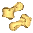
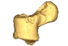
M3#336The specimen FSL848325 is separated in two fragments: the anterior part bears the incisors, the deciduous and permanent canines, while the posterior part bears the right P3, P4, M1 and M2. The P2 is isolated. When combined, the cranium length is approximatively 10.5 cm long. The anterior part is 6.9 cm long and 2.15 cm wide (taken at the level of the P1). The posterior part is 4.8 cm long. The anterior part of the cranium is very narrow. Type: "3D_surfaces"doi: 10.18563/m3.sf.336 state:published |
Download 3D surface file |
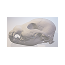
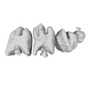
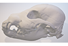
This contribution comprises the 3D models of three wolf pup skulls, which were used for the publication by Geiger et al. 2017 on Neomorphosis and heterochrony of skull shape in dog domestication.
Canis lupus CLL2 View specimen
|
M3#3123d model of a wolf pup skull Type: "3D_surfaces"doi: 10.18563/m3.sf.312 state:published |
Download 3D surface file |
Canis lupus CLL4 View specimen
|
M3#3133d model of a wolf pup skull Type: "3D_surfaces"doi: 10.18563/m3.sf.313 state:published |
Download 3D surface file |
Canis lupus CLL5 View specimen
|
M3#3143d model of a wolf pup skull Type: "3D_surfaces"doi: 10.18563/m3.sf.314 state:published |
Download 3D surface file |
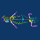
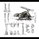
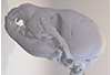
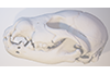
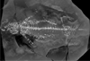
The present contribution contains the 3D model and dataset analyzed in the following publication: Scheyer, T. M., J. M. Neenan, T. Bodogan, H. Furrer, C. Obrist, and M. Plamondon. 2017. A new, exceptionally preserved juvenile specimen of Eusaurosphargis dalsassoi (Diapsida) and implications for Mesozoic marine diapsid phylogeny. Scientific Reports, https://doi.org/10.1038/s41598-017-04514-x .
Eusaurosphargis dalsassoi PIMUZ A/III 4380 View specimen
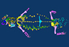
|
M3#17994 extracted surfaces of skeletal elements of PIMUZ A/III 4380 Type: "3D_surfaces"doi: 10.18563/m3.sf.179 state:published |
Download 3D surface file |
|
M3#180Accompanying CT scan dataset Type: "3D_CT"doi: 10.18563/m3.sf.180 state:published |
Download CT data |


