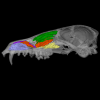
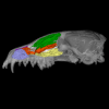
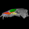
Explodable 3D Dog Skull for Veterinary Education
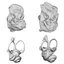
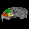
3D models of Ocnotherium skull
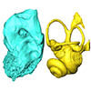
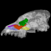
3D models of Kalakocetus, the earliest Cetacea
3D GM dataset of bird skeletal variation
Skeletal embryonic development in the catshark
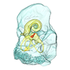
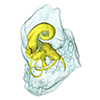
Bony connexions of the petrosal bone of extant hippos
bony labyrinth (14) , inner ear (11) , geometric morphometrics (10) , CT-scan (10) , Eocene (10) , Micro-CT (9) , Miocene (8)
Lionel Hautier (24) , Maëva Judith Orliac (23) , Laurent Marivaux (18) , Renaud Lebrun (14) , Rodolphe Tabuce (14) , Pierre-Olivier Antoine (13) , Bastien Mennecart (13)

|
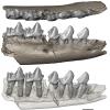
3D models related to the publication: "From teeth to pad: tooth loss and development of keratinous structures in sirenians"Lionel Hautier
Published online: 29/11/2023 |
|
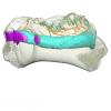
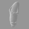
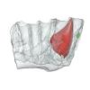
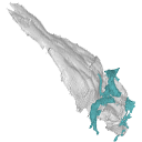
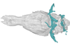
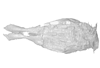
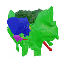
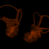
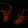
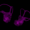
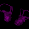
M3#1275Internal mandibular morphology. Orange = dorsal canaliculi; purple = mental branches; cyan = mandibular canal; dark blue = teeth; green = tooth alveoli. Type: "3D_surfaces"doi: 10.18563/m3.sf.1275 state:published |
Download 3D surface file |
Dugong dugon 2023.66 View specimen
|
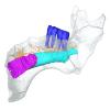
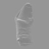
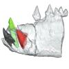
M3#1274Internal mandibular morphology. Orange = dorsal canaliculi; purple = mental branches; cyan = mandibular canal; dark blue = teeth; green = tooth alveoli. Type: "3D_surfaces"doi: 10.18563/m3.sf.1274 state:published |
Download 3D surface file |
Dugong dugon 5386 View specimen
|
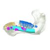
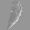
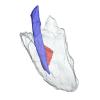
M3#1276Internal mandibular morphology. Orange = dorsal canaliculi; purple = mental branches; cyan = mandibular canal; dark blue = teeth; green = tooth alveoli. Type: "3D_surfaces"doi: 10.18563/m3.sf.1276 state:published |
Download 3D surface file |
Dugong dugon 1848.8.29.7/GERM 1027g View specimen
|
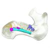
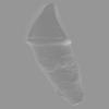
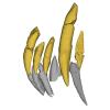
M3#1277Internal mandibular morphology. Orange = dorsal canaliculi; purple = mental branches; cyan = mandibular canal; dark blue = teeth. Type: "3D_surfaces"doi: 10.18563/m3.sf.1277 state:published |
Download 3D surface file |
Dugong dugon 1991.413 View specimen
|
M3#1278Internal mandibular morphology. Orange = dorsal canaliculi; purple = mental branches; cyan = mandibular canal; dark blue = teeth. Type: "3D_surfaces"doi: 10.18563/m3.sf.1278 state:published |
Download 3D surface file |
Dugong dugon 1991.427 View specimen
|
M3#1279Internal mandibular morphology. Orange = dorsal canaliculi; purple = mental branches; cyan = mandibular canal; dark blue = teeth. Type: "3D_surfaces"doi: 10.18563/m3.sf.1279 state:published |
Download 3D surface file |
Dugong dugon 2017-3-9 View specimen
|
M3#1280Internal mandibular morphology. Orange = dorsal canaliculi; purple = mental branches; cyan = mandibular canal; dark blue = teeth. Type: "3D_surfaces"doi: 10.18563/m3.sf.1280 state:published |
Download 3D surface file |
Eosiren lybica 1913-22 View specimen
|
M3#1281Internal mandibular morphology. Orange = dorsal canaliculi; purple = mental branches; cyan = mandibular canal; dark blue = teeth; green = tooth alveoli. Type: "3D_surfaces"doi: 10.18563/m3.sf.1281 state:published |
Download 3D surface file |
Halitherium taulannense RGHP C001 View specimen
|
M3#1282Internal mandibular morphology. Cyan = mandibular canal; dark blue = teeth. Type: "3D_surfaces"doi: 10.18563/m3.sf.1282 state:published |
Download 3D surface file |
Halitherium taulannense RGHP C009 View specimen
|
M3#1283Internal mandibular morphology. Orange = dorsal canaliculi; purple = mental branches; cyan = mandibular canal; dark blue = teeth; green = tooth alveoli. Type: "3D_surfaces"doi: 10.18563/m3.sf.1283 state:published |
Download 3D surface file |
Hydrodamalis gigas 1947.10.21.1 View specimen
|
M3#1284Internal mandibular morphology. Orange = dorsal canaliculi; purple = mental branches; cyan = mandibular canal Type: "3D_surfaces"doi: 10.18563/m3.sf.1284 state:published |
Download 3D surface file |
Hydrodamalis gigas C1021 View specimen

|
M3#1285Anterior part of the mandible Type: "3D_surfaces"doi: 10.18563/m3.sf.1285 state:published |
Download 3D surface file |
|
M3#1286Posterior part of the mandible Type: "3D_surfaces"doi: 10.18563/m3.sf.1286 state:published |
Download 3D surface file |
Hydrodamalis gigas 2023.67 View specimen
|
M3#1287Internal mandibular morphology. Orange = dorsal canaliculi; purple = mental branches; cyan = mandibular canal Type: "3D_surfaces"doi: 10.18563/m3.sf.1287 state:published |
Download 3D surface file |
Libysiren sickenbergi M.82429 View specimen
|
M3#1288Internal mandibular morphology. Orange = dorsal canaliculi; purple = mental branches; cyan = mandibular canal; dark blue = teeth; green = tooth alveoli. Type: "3D_surfaces"doi: 10.18563/m3.sf.1288 state:published |
Download 3D surface file |
Libysiren sickenbergi M.45675 View specimen
|
M3#1289Internal mandibular morphology. Orange = dorsal canaliculi; purple = mental branches; cyan = mandibular canal; dark blue = teeth; green = tooth alveoli. Type: "3D_surfaces"doi: 10.18563/m3.sf.1289 state:published |
Download 3D surface file |
Prorastomus sirenoides OR.448976 View specimen

|
M3#1290Internal morphology of the left mandible. Orange = dorsal canaliculi; purple = mental branches; cyan = mandibular canal; dark blue = teeth; green = tooth alveoli. Type: "3D_surfaces"doi: 10.18563/m3.sf.1290 state:published |
Download 3D surface file |
|
M3#1304Internal morphology of the right mandible. Orange = dorsal canaliculi; purple = mental branches; cyan = mandibular canal; dark blue = teeth. Type: "3D_surfaces"doi: 10.18563/m3.sf.1304 state:published |
Download 3D surface file |
Ribodon limbatus M.7073 View specimen
|
M3#1292Internal mandibular morphology. Orange = dorsal canaliculi; purple = mental branches; cyan = mandibular canal; dark blue = teeth; green = tooth alveoli. Type: "3D_surfaces"doi: 10.18563/m3.sf.1292 state:published |
Download 3D surface file |
Rytiodus capgrandi PAL2017-8-1 View specimen
|
M3#1293Internal mandibular morphology. Orange = dorsal canaliculi; purple = mental branches; cyan = mandibular canal; dark blue = teeth; green = tooth alveoli. Type: "3D_surfaces"doi: 10.18563/m3.sf.1293 state:published |
Download 3D surface file |
Trichechus inunguis 1868.12.19.2 View specimen
|
M3#1294Internal mandibular morphology. Orange = dorsal canaliculi; purple = mental branches; cyan = mandibular canal; dark blue = teeth; green = tooth alveoli. Type: "3D_surfaces"doi: 10.18563/m3.sf.1294 state:published |
Download 3D surface file |
Trichechus manatus 1843.3.10.12 View specimen
|
M3#1295Internal mandibular morphology. Orange = dorsal canaliculi; purple = mental branches; cyan = mandibular canal; dark blue = teeth. Type: "3D_surfaces"doi: 10.18563/m3.sf.1295 state:published |
Download 3D surface file |
Trichechus manatus 1864.6.5.1 View specimen
|
M3#1296Internal mandibular morphology. Orange = dorsal canaliculi; purple = mental branches; cyan = mandibular canal; dark blue = teeth; green = tooth alveoli. Type: "3D_surfaces"doi: 10.18563/m3.sf.1296 state:published |
Download 3D surface file |
Trichechus manatus 1950.1.23.1 View specimen
|
M3#1297Internal mandibular morphology. Orange = dorsal canaliculi; purple = mental branches; cyan = mandibular canal; dark blue = teeth; green = tooth alveoli. Type: "3D_surfaces"doi: 10.18563/m3.sf.1297 state:published |
Download 3D surface file |
Trichechus senegalensis 1885.6.30.2 View specimen
|
M3#1298Internal mandibular morphology. Orange = dorsal canaliculi; purple = mental branches; cyan = mandibular canal; dark blue = teeth; green = tooth alveoli. Type: "3D_surfaces"doi: 10.18563/m3.sf.1298 state:published |
Download 3D surface file |
Trichechus senegalensis 1894.7.25.8 View specimen
|
M3#1299Internal mandibular morphology. Orange = dorsal canaliculi; purple = mental branches; cyan = mandibular canal; dark blue = teeth. Type: "3D_surfaces"doi: 10.18563/m3.sf.1299 state:published |
Download 3D surface file |
Trichechus senegalensis V97 View specimen
|
M3#1302Mandibular internal morphology. Orange = dorsal canaliculi; purple = mental branches; cyan = mandibular canal; dark blue = teeth. Type: "3D_surfaces"doi: 10.18563/m3.sf.1302 state:published |
Download 3D surface file |
Trichechus sp. 65.4.28.9 View specimen
|
M3#1300Internal mandibular morphology. Orange = dorsal canaliculi; purple = mental branches; cyan = mandibular canal; dark blue = teeth; green = tooth alveoli. Type: "3D_surfaces"doi: 10.18563/m3.sf.1300 state:published |
Download 3D surface file |
Dugong dugon 1946.8.6.2 View specimen
|
M3#1301Mandibular internal morphology. Orange = dorsal canaliculi; purple = mental branches; cyan = mandibular canal; dark blue = teeth. Type: "3D_surfaces"doi: 10.18563/m3.sf.1301 state:published |
Download 3D surface file |
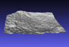
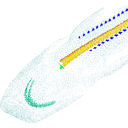
We provide a 3D reconstruction of the skull of Latimeria chalumnae that can be easily accessed and visualized for a better understanding of its cranial anatomy. Different skeletal elements are saved as separate PLY files that can be combined to visualize the entire skull or isolated to virtually dissect the skull. We included some guidelines for a fast and easy visualization of the 3D skull.
Latimeria chalumnae MHNG 1080.070 View specimen

|
M3#1254the skeletal elements of the skull of Latimeria chalumnae included in 26 different PLY files Type: "3D_surfaces"doi: 10.18563/m3.sf.1254 state:published |
Download 3D surface file |
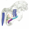
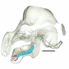
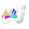
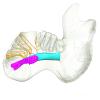
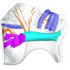
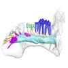
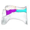
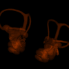
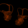
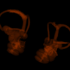
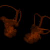
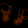
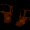
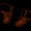
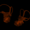
The present 3D Dataset contains the 3D models analyzed in Mennecart, B., Duranthon, F., & Costeur, L. 2024. Systematic contribution of the auditory region to the knowledge of the oldest European Bovidae (Mammalia, Ruminantia). Journal of Anatomy XXX. https://doi.org/10.1111/joa.14132
Pusillutragus montrealensis MHNT.PAL.2015.0.2261.4 View specimen
|
M3#1522Right petrosal, bony labyrinth, stapes Type: "3D_surfaces"doi: 10.18563/m3.sf.1522 state:published |
Download 3D surface file |
Pusillutragus montrealensis MHNT.PAL.2015.0.2261.9 View specimen

|
M3#1523Left petrosal and left bony labyrinth Type: "3D_surfaces"doi: 10.18563/m3.sf.1523 state:published |
Download 3D surface file |
Eotragus artenensis SMNS-P-41625 View specimen
|
M3#1524Petrosal (right), bony labyrinth (left) Type: "3D_surfaces"doi: 10.18563/m3.sf.1524 state:published |
Download 3D surface file |
Eotragus clavatus NMB San.15056 View specimen
|
M3#1528Right petrosal and right bony labyrinth Type: "3D_surfaces"doi: 10.18563/m3.sf.1528 state:published |
Download 3D surface file |
Eotragus clavatus NMB San.15055 View specimen
|
M3#1526Left Petrosal Type: "3D_surfaces"doi: 10.18563/m3.sf.1526 state:published |
Download 3D surface file |
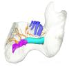
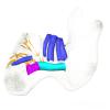
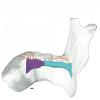
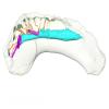
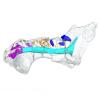
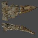
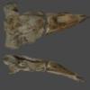
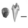
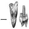
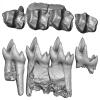
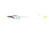
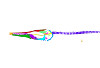
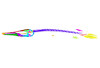
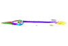
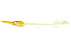
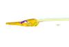
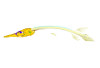
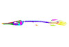
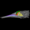
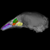
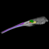
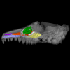
The present 3D dataset contains 3D models of new material from the middle Eocene of the Upper Subathu Formation in the Kalakot area (India), documenting the anterior dentition of the raoellid Indohyus indirae. Raoellidae are closely related to stem cetaceans and bring crucial information to understand the earliest phase of land to water transition in Cetacea.
Indohyus indirae GU/RJ/31 View specimen
|
M3#1505Right i1 Type: "3D_surfaces"doi: 10.18563/m3.sf.1505 state:published |
Download 3D surface file |
Indohyus indirae GU/RJ/32 View specimen
|
M3#1506Right i1 Type: "3D_surfaces"doi: 10.18563/m3.sf.1506 state:published |
Download 3D surface file |
Indohyus indirae GU/RJ/16 View specimen
|
M3#1507Left I3 Type: "3D_surfaces"doi: 10.18563/m3.sf.1507 state:published |
Download 3D surface file |
Indohyus indirae GU/RJ/23 View specimen
|
M3#1508Left I2 Type: "3D_surfaces"doi: 10.18563/m3.sf.1508 state:published |
Download 3D surface file |
Indohyus indirae GU/RJ/25 View specimen
|
M3#1509Left I1 Type: "3D_surfaces"doi: 10.18563/m3.sf.1509 state:published |
Download 3D surface file |
Indohyus indirae GU/RJ/26 View specimen
|
M3#1510Right I3 Type: "3D_surfaces"doi: 10.18563/m3.sf.1510 state:published |
Download 3D surface file |
Indohyus indirae GU/RJ/57 View specimen
|
M3#1511Left I1 Type: "3D_surfaces"doi: 10.18563/m3.sf.1511 state:published |
Download 3D surface file |
Indohyus indirae GU/RJ/61 View specimen
|
M3#1512Right upper canine Type: "3D_surfaces"doi: 10.18563/m3.sf.1512 state:published |
Download 3D surface file |
Indohyus indirae GU/RJ/63 View specimen
|
M3#1513Left upper canine Type: "3D_surfaces"doi: 10.18563/m3.sf.1513 state:published |
Download 3D surface file |
Indohyus indirae GU/RJ/74 View specimen
|
M3#1514Left upper canine Type: "3D_surfaces"doi: 10.18563/m3.sf.1514 state:published |
Download 3D surface file |
Indohyus indirae GU/RJ/439 View specimen
|
M3#1515Left upper canine Type: "3D_surfaces"doi: 10.18563/m3.sf.1515 state:published |
Download 3D surface file |
Indohyus indirae GU/RJ/457 View specimen
|
M3#1516Left upper canine Type: "3D_surfaces"doi: 10.18563/m3.sf.1516 state:published |
Download 3D surface file |
Indohyus indirae GU/RJ/846 View specimen
|
M3#1517Left upper canine Type: "3D_surfaces"doi: 10.18563/m3.sf.1517 state:published |
Download 3D surface file |
Indohyus indirae GU/RJ/822 View specimen
|
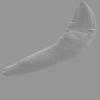
M3#1518Right fragmentary maxillary with decidual canine and I3 Type: "3D_surfaces"doi: 10.18563/m3.sf.1518 state:published |
Download 3D surface file |
Indohyus indirae GU/RJ/824 View specimen
|
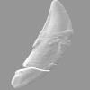
M3#1519Right fragmentary mandible with lower canine and small part of the i3 Type: "3D_surfaces"doi: 10.18563/m3.sf.1519 state:published |
Download 3D surface file |
Indohyus indirae GU/RJ/838 View specimen
|
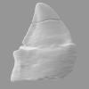
M3#1520Right fragmentary mandible with permanent i2, i3 and canine and small part of the root of the decidual i3 Type: "3D_surfaces"doi: 10.18563/m3.sf.1520 state:published |
Download 3D surface file |
Indohyus indirae GU/RJ/842 View specimen
|
M3#1521Left fragmentary mandible with decidual and permanent canine Type: "3D_surfaces"doi: 10.18563/m3.sf.1521 state:published |
Download 3D surface file |
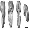
Indohyus indirae GU/RJ/26,32,56,57,457,822,838,842 : Composite Anterior dentition View specimen
|
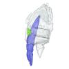
M3#15293D composite reconstruction of the anterior dentition of Indohyus indirae with GU/RJ/57 (I1), 56 (I2), 26 (I3), 457 (Upper canine), 822 (P1), 32 (i1), 838 (i2, i3 and lower canine) and 842 (p1) Type: "3D_surfaces"doi: 10.18563/m3.sf.1529 state:published |
Download 3D surface file |
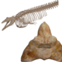
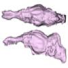
The present 3D Dataset contains the 3D models analyzed in Bianucci et al. 2023, A heavyweight early whale pushes the boundaries of vertebrate morphology, Nature. These include bones of the holotype of new species Perucetus colossus (MUSM 3248), as well as the articulated skeleton of Cynthiacetus peruvianus (holotype, MNHN.F.PRU10). The latter was used to estimate the total skeleton volume of P. colossus.
Perucetus colossus MUSM 3248 View specimen

|
M3#1131Thirteen vertebrae, rib, and innominate of Perucetus colossus (holotype, MUSM NNNN). Type: "3D_surfaces"doi: 10.18563/m3.sf.1131 state:published |
Download 3D surface file |
Cynthiacetus peruvianus MNHN.F.PRU10 View specimen
|
M3#1130Articulated skeleton of the holotype of Cynthiacetus peruvianus MNHN.F.PRU10 Type: "3D_surfaces"doi: 10.18563/m3.sf.1130 state:published |
Download 3D surface file |
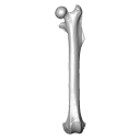
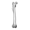
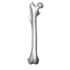
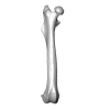
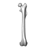
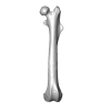
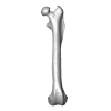
This 3D Dataset includes the 3D models analysed in Wölfer J & Hautier L. 2024 Inferring the locomotor ecology of two of the oldest fossil squirrels: influence of operationalisation, trait, body size, and machine learning method. Proceedings of the Royal Society B. https://doi.org/10.1098/rspb.2024-0743
Palaeosciurus goti MGB125 View specimen

|
M3#1577Left femur of Palaeosciurus goti Type: "3D_surfaces"doi: 10.18563/m3.sf.1577 state:published |
Download 3D surface file |
Palaeosciurus feignouxi GER291 View specimen
|
M3#1578Right femur of Palaeosciurus feignouxi Type: "3D_surfaces"doi: 10.18563/m3.sf.1578 state:published |
Download 3D surface file |
Palaeosciurus feignouxi GER293 View specimen
|
M3#1579Right femur of Palaeosciurus feignouxi Type: "3D_surfaces"doi: 10.18563/m3.sf.1579 state:published |
Download 3D surface file |
Palaeosciurus feignouxi GER294 View specimen
|
M3#1580Right femur of Palaeosciurus feignouxi Type: "3D_surfaces"doi: 10.18563/m3.sf.1580 state:published |
Download 3D surface file |
Palaeosciurus feignouxi GER296 View specimen
|
M3#1581Left femur of Palaeosciurus feignouxi Type: "3D_surfaces"doi: 10.18563/m3.sf.1581 state:published |
Download 3D surface file |
Palaeosciurus feignouxi GER298 View specimen
|
M3#1582Left femur of Palaeosciurus feignouxi Type: "3D_surfaces"doi: 10.18563/m3.sf.1582 state:published |
Download 3D surface file |
Palaeosciurus feignouxi GER299 View specimen
|
M3#1583Left femur of Palaeosciurus feignouxi Type: "3D_surfaces"doi: 10.18563/m3.sf.1583 state:published |
Download 3D surface file |
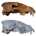
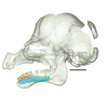
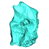
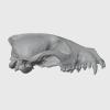
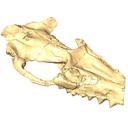
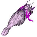
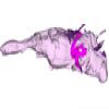
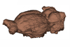
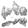
Speothos pacivorus is an extinct South American canid (Canidae: Cerdocyonina) from the Pleistocene of Lagoa Santa Karst, Central Brazil. This taxon is one of the hypercarnivore canids that vanished from the continent at the end of Pleistocene. Although all remains of Speothos pacivorus were collected in the 19th century by the Danish naturalist Peter W. Lund, few studies have committed to an in-depth analysis of the taxon and the known specimens. Here, we analyzed all biological remains of S. pacivorus hosted in the Peter Lund/Quaternary Collection at the Natural History Museum of Denmark, Copenhagen, by listing and illustrating all its specimens known to date. We also conducted a reconstruction of the holotype, an almost complete cranium, based on a µCT scan, producing an undeformed and crack-free three-dimensional model. With this data available we aim to foster new research on this elusive species.
Speothos pacivorus NHMD:211341 View specimen
|
M3#1475Holotype of Speothos pacivorus Type: "3D_surfaces"doi: 10.18563/m3.sf.1475 state:published |
Download 3D surface file |
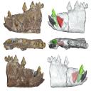
The present 3D Dataset contains the 3D model of the skin of Allosaurus described in Hendrickx, C. et al. in press. Morphology and distribution of scales, dermal ossifications, and other non-feather integumentary structures in non-avialan theropod dinosaurs. Biological Reviews.
Allosaurus jimmadseni UMNH VP C481 View specimen
|
M3#902The material consists of a 3D reconstruction of the counterpart of a 30 cm2 patch of skin impression associated with the anterior dorsal ribs/pectoral region of the specimen of Allosaurus jimmadseni UMNH VP C481. The skin shows a semi-uniform basement of 1-2 mm diameter pebbles with a smaller number of slightly larger (up to 3 mm) ovoid scales. The irregular shape, distribution, and overall small size of these larger scales suggest that they are not classifiable as feature scales but rather as variations in the basement scales. Type: "3D_surfaces"doi: 10.18563/m3.sf.902 state:published |
Download 3D surface file |
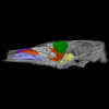
The present 3D Dataset contains the 3D models of the endocranial cast of two specimens of Indohyus indirae described in the article entitled “The endocranial cast of Indohyus (Artiodactyla, Raoellidae): the origin of the cetacean brain” (Orliac and Thewissen, 2021). They represent the cast of the main cavity of the braincase as well as associated intraosseous sinuses.
Indohyus indirae RR 207 View specimen
|
M3#710cast of the main endocranial cavity and associated intraosseous sinuses Type: "3D_surfaces"doi: 10.18563/m3.sf.710 state:published |
Download 3D surface file |
Indohyus indirae RR 601 View specimen
|
M3#711casts of the main endocranial cavity Type: "3D_surfaces"doi: 10.18563/m3.sf.711 state:published |
Download 3D surface file |
Current knowledge on the skeletogenesis of Chondrichthyes is scarce compared with their extant sister group, the bony fishes. Most of the previously described developmental tables in Chondrichthyes have focused on embryonic external morphology only. Due to its small body size and relative simplicity to raise eggs in laboratory conditions, the small-spotted catshark Scyliorhinus canicula has emerged as a reference species to describe developmental mechanisms in the Chondrichthyes lineage. Here we investigate the dynamic of mineralization in a set of six embryonic specimens using X-ray microtomography and describe the developing units of both the dermal skeleton (teeth and dermal scales) and endoskeleton (vertebral axis). This preliminary data on skeletogenesis in the catshark sets the first bases to a more complete investigation of the skeletal developmental in Chondrichthyes. It should provide comparison points with data known in osteichthyans and could thus be used in the broader context of gnathostome skeletal evolution.
Scyliorhinus canicula SC6_2_2015_03_20 View specimen

|
M3#50Mineralized skeleton of a 6,2 cm long embryo of Scyliorhinus canicula Type: "3D_surfaces"doi: 10.18563/m3.sf.50 state:published |
Download 3D surface file |
Scyliorhinus canicula SC6_7_2015_03_20 View specimen

|
M3#51Mineralized skeleton of a 6,7 cm long embryo of Scyliorhinus canicula Type: "3D_surfaces"doi: 10.18563/m3.sf.51 state:published |
Download 3D surface file |
Scyliorhinus canicula SC7_1_2015_04_03 View specimen

|
M3#52Mineralized skeleton of a 7,1 cm long embryo of Scyliorhinus canicula Type: "3D_surfaces"doi: 10.18563/m3.sf.52 state:published |
Download 3D surface file |
Scyliorhinus canicula SC7_5_2015_03_13 View specimen

|
M3#53Mineralized skeleton of a 7,5 cm long embryo of Scyliorhinus canicula Type: "3D_surfaces"doi: 10.18563/m3.sf.53 state:published |
Download 3D surface file |
Scyliorhinus canicula SC8_2015_03_20 View specimen

|
M3#54Mineralized skeleton of a 8 cm long embryo of Scyliorhinus canicula Type: "3D_surfaces"doi: 10.18563/m3.sf.54 state:published |
Download 3D surface file |
Scyliorhinus canicula SC10_2015_02_27 View specimen

|
M3#55Mineralized skeleton of a 10 cm long embryo of Scyliorhinus canicula Type: "3D_surfaces"doi: 10.18563/m3.sf.55 state:published |
Download 3D surface file |
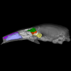
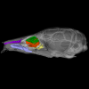
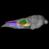
The present 3D Dataset contains the 3D model of the skull of the raoellid Indohyus indirae described in Patel et al. 2024.
Indohyus indirae RR 207 View specimen
|
M3#1259dorsoventrally crushed skull Type: "3D_surfaces"doi: 10.18563/m3.sf.1259 state:published |
Download 3D surface file |
Indohyus indirae RR 601 View specimen
|
M3#1268dorsoventrally crushed skull Type: "3D_surfaces"doi: 10.18563/m3.sf.1268 state:published |
Download 3D surface file |
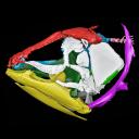
The present 3D Dataset contains the 3D models analyzed in Pochat-Cottilloux Y., Martin J.E., Jouve S., Perrichon G., Adrien J., Salaviale C., de Muizon C., Cespedes R. & Amiot R. (2021). The neuroanatomy of Zulmasuchus querejazus (Crocodylomorpha, Sebecidae) and its implications for the paleoecology of sebecosuchians. The Anatomical Record, https://doi.org/10.1002/ar.24826
Zulmasuchus querejazus MHNC 6672 View specimen

|
M3#798Left endosseous labyrinth of Z. querejazus (MHNC 6672). Type: "3D_surfaces"doi: 10.18563/m3.sf.798 state:published |
Download 3D surface file |

|
M3#799Reconstruction of the endocranial cavities of Z. querejazus (MHNC 6672). Type: "3D_surfaces"doi: 10.18563/m3.sf.799 state:published |
Download 3D surface file |
|
M3#800Three-dimensional reconstruction of the pneumatic cavities within the braincase of Z. querejazus (MHNC 6672) Type: "3D_surfaces"doi: 10.18563/m3.sf.800 state:published |
Download 3D surface file |
This contribution contains the 3D model described and figured in the following publication: Albino, A., Carrillo-Briceño, J. D. & Neenan, J. M. 2016. An enigmatic aquatic snake from the Cenomanian of northern South America. PeerJ 4:e2027 http://dx.doi.org/10.7717/peerj.2027
Lunaophis aquaticus MCNC-1827-F View specimen
|
M3#116Articulated precloacal vertebrae of Lunaophis aquaticus Type: "3D_surfaces"doi: 10.18563/m3.sf.116 state:published |
Download 3D surface file |
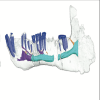
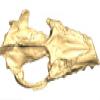
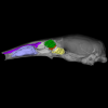
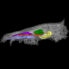
The present 3D dataset contains 3D models of the endocranial cast of the raoellid Khirtharia inflata retrieved from the middle Eocene of the Upper Subathu Formation in the Kalakot area (India). Raoellidae are closely related to stem cetaceans and bring crucial information to understand the earliest phase of land to water transition in Cetacea.
Khirtharia inflata GU/RJ/197 View specimen
|
M3#1608labeled cast of the endocranial cavity Type: "3D_surfaces"doi: 10.18563/m3.sf.1608 state:published |
Download 3D surface file |
|
M3#1609endocast and associated sinuses Type: "3D_surfaces"doi: 10.18563/m3.sf.1609 state:published |
Download 3D surface file |
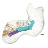
Our knowledge of the external brain morphology of the late Eocene artiodactyl ungulate Mixtotherium, relies on a plaster model realized on a specimen from the Victor Brun Museum in Montauban (France) and described by Dechaseaux (1973). Here, based on micro CT-scan data, we virtually reconstruct the 3D cast of the empty cavity of the partial cranium MA PHQ 716 from the Victor Brun Museum and compare it to the plaster model illustrated and described by Dechaseaux (1973). Indeed, the specimen from which the original plaster endocast originates was not identified by Dechaseaux by a specimen number. We confirm here that the studied specimen was indeed the one described and illustrated by Dechaseaux (1973). We also reconstruct a second, more detailed, model providing additional morphological and quantitative observations made available by micro CT scan investigation such as precisions on the neopallium folding and endocranial volumes.
Mixtotherium cuspidatum MA PHQ 716 View specimen
|
M3#857endocast of the brain cavity Type: "3D_surfaces"doi: 10.18563/m3.sf.857 state:published |
Download 3D surface file |
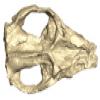
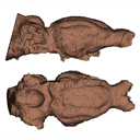
The present 3D Dataset contains the 3D models of the skull of the holotype of Miocaperea pulchra.
Miocaperea pulchra SMNS-P-46978 View specimen
|
M3#1656Blender file containing two models (the skull being preserved in two parts) Type: "3D_surfaces"doi: 10.18563/m3.sf.1656 state:published |
Download 3D surface file |
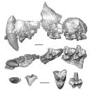
This contribution contains the three-dimensional models of the most complete and/or informative fossil materials attributed to Peradectes crocheti Gernelle, 2024, the earliest peradectid metatherian species of Europe, from its type locality (Palette, Provence, ~55 Ma). These specimens were analyzed and discussed in: Gernelle et al. (2024), Taxonomy and evolutionary history of peradectids (Metatheria): new data from the early Eocene of France. https://doi.org/10.1007/s10914-024-09724-5
Peradectes crocheti MHN.AIX.PV.2018.26.14 View specimen
|
M3#14993D surface model of MHN.AIX.PV.2018.26.14, fragmentary left maxilla with C-P1, anterior root of P2, and M1-M3 Type: "3D_surfaces"doi: 10.18563/m3.sf.1499 state:published |
Download 3D surface file |
Peradectes crocheti MHN.AIX.PV.2017.6.6 View specimen
|
M3#15003D surface model of MHN.AIX.PV.2017.6.6, left P2 Type: "3D_surfaces"doi: 10.18563/m3.sf.1500 state:published |
Download 3D surface file |
Peradectes crocheti MHN.AIX.PV.2017.6.7 View specimen
|
M3#15013D surface model of MHN.AIX.PV.2017.6.7, left M3 Type: "3D_surfaces"doi: 10.18563/m3.sf.1501 state:published |
Download 3D surface file |
Peradectes crocheti MHN.AIX.PV.2017.6.8 View specimen
|
M3#15023D surface model of MHN.AIX.PV.2017.6.8, right hemi-mandible fragment with canine alveolus, posterior root of p1, partial p2, p3, partial m1, and m2-m3 Type: "3D_surfaces"doi: 10.18563/m3.sf.1502 state:published |
Download 3D surface file |
Peradectes crocheti MHN.AIX.PV.2017.6.9 View specimen
|
M3#15033D surface model of MHN.AIX.PV.2017.6.9, leftm1-m4 row with fragments of dentary Type: "3D_surfaces"doi: 10.18563/m3.sf.1503 state:published |
Download 3D surface file |
Peradectes crocheti MHN.AIX.PV.2017.6.14 View specimen
|
M3#15043D surface model of MHN.AIX.PV.2017.6.14, right astragalus Type: "3D_surfaces"doi: 10.18563/m3.sf.1504 state:published |
Download 3D surface file |
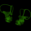
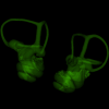
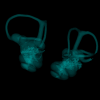
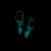
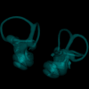
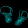
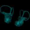
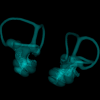
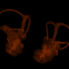
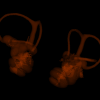
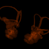
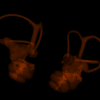
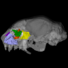
This contribution contains 3D models of left and right house mouse (Mus musculus domesticus) inner ears analyzed in Renaud et al. (2024). The studied mice belong to four groups: wild-trapped mice, wild-derived lab offspring, a typical laboratory strain (Swiss) and hybrids between wild-derived and Swiss mice. They have been analyzed to assess the impact of mobility reduction on inner ear morphology, including patterns of divergence, levels of inter-individual variance (disparity) and intra-individual variance (fluctuating asymmetry)
Mus musculus Tourch_7819 View specimen
|
M3#1366Two bony labyrinths Type: "3D_surfaces"doi: 10.18563/m3.sf.1366 state:published |
Download 3D surface file |
Mus musculus Tourch_7821 View specimen
|
M3#1367Two bony labyrinths Type: "3D_surfaces"doi: 10.18563/m3.sf.1367 state:published |
Download 3D surface file |
Mus musculus Tourch_7839 View specimen

|
M3#1368Two bony labyrinths Type: "3D_surfaces"doi: 10.18563/m3.sf.1368 state:published |
Download 3D surface file |
Mus musculus Tourch_7873 View specimen

|
M3#1369Two bony labyrinths Type: "3D_surfaces"doi: 10.18563/m3.sf.1369 state:published |
Download 3D surface file |
Mus musculus Tourch_7877 View specimen

|
M3#1370Two bony labyrinths Type: "3D_surfaces"doi: 10.18563/m3.sf.1370 state:published |
Download 3D surface file |
Mus musculus Tourch_7922 View specimen
|
M3#1371Two bony labyrinths Type: "3D_surfaces"doi: 10.18563/m3.sf.1371 state:published |
Download 3D surface file |
Mus musculus Tourch_7923 View specimen
|
M3#1372Two bony labyrinths Type: "3D_surfaces"doi: 10.18563/m3.sf.1372 state:published |
Download 3D surface file |
Mus musculus Tourch_7925 View specimen
|
M3#1373Two bony labyrinths Type: "3D_surfaces"doi: 10.18563/m3.sf.1373 state:published |
Download 3D surface file |
Mus musculus Tourch_7927 View specimen
|
M3#1374Two bony labyrinths Type: "3D_surfaces"doi: 10.18563/m3.sf.1374 state:published |
Download 3D surface file |
Mus musculus Tourch_7932 View specimen
|
M3#1375Two bony labyrinths Type: "3D_surfaces"doi: 10.18563/m3.sf.1375 state:published |
Download 3D surface file |
Mus musculus Bal02 View specimen
|
M3#1320Two bony labyrinths Type: "3D_surfaces"doi: 10.18563/m3.sf.1320 state:published |
Download 3D surface file |
Mus musculus Bal04 View specimen
|
M3#1321Two bony labyrinths Type: "3D_surfaces"doi: 10.18563/m3.sf.1321 state:published |
Download 3D surface file |
Mus musculus Bal06 View specimen
|
M3#1322Two bony labyrinths Type: "3D_surfaces"doi: 10.18563/m3.sf.1322 state:published |
Download 3D surface file |
Mus musculus Bal08 View specimen

|
M3#1323Two bony labyrinths Type: "3D_surfaces"doi: 10.18563/m3.sf.1323 state:published |
Download 3D surface file |
Mus musculus Bal11 View specimen
|
M3#1324Two bony labyrinths Type: "3D_surfaces"doi: 10.18563/m3.sf.1324 state:published |
Download 3D surface file |
Mus musculus Bal12 View specimen
|
M3#1325Two bony labyrinths Type: "3D_surfaces"doi: 10.18563/m3.sf.1325 state:published |
Download 3D surface file |
Mus musculus Bal15 View specimen

|
M3#1326Two bony labyrinths Type: "3D_surfaces"doi: 10.18563/m3.sf.1326 state:published |
Download 3D surface file |
Mus musculus Bal16 View specimen
|
M3#1327Two bony labyrinths Type: "3D_surfaces"doi: 10.18563/m3.sf.1327 state:published |
Download 3D surface file |
Mus musculus Bal17 View specimen
|
M3#1328Two bony labyrinths Type: "3D_surfaces"doi: 10.18563/m3.sf.1328 state:published |
Download 3D surface file |
Mus musculus Bal18 View specimen
|
M3#1329Two bony labyrinths Type: "3D_surfaces"doi: 10.18563/m3.sf.1329 state:published |
Download 3D surface file |
Mus musculus Bal19 View specimen
|
M3#1330Two bony labyrinths Type: "3D_surfaces"doi: 10.18563/m3.sf.1330 state:published |
Download 3D surface file |
Mus musculus Bal20 View specimen

|
M3#1331Two bony labyrinths Type: "3D_surfaces"doi: 10.18563/m3.sf.1331 state:published |
Download 3D surface file |
Mus musculus Bal21 View specimen
|
M3#1332Two bony labyrinths Type: "3D_surfaces"doi: 10.18563/m3.sf.1332 state:published |
Download 3D surface file |
Mus musculus Bal22 View specimen
|
M3#1333Two bony labyrinths Type: "3D_surfaces"doi: 10.18563/m3.sf.1333 state:published |
Download 3D surface file |
Mus musculus Bal23 View specimen
|
M3#1334Two bony labyrinths Type: "3D_surfaces"doi: 10.18563/m3.sf.1334 state:published |
Download 3D surface file |
Mus musculus Bal24 View specimen
|
M3#1335Two bony labyrinths Type: "3D_surfaces"doi: 10.18563/m3.sf.1335 state:published |
Download 3D surface file |
Mus musculus Bal25 View specimen
|
M3#1336Two bony labyrinths Type: "3D_surfaces"doi: 10.18563/m3.sf.1336 state:published |
Download 3D surface file |
Mus musculus Balan_LAB_035 View specimen
|
M3#1337Two bony labyrinths Type: "3D_surfaces"doi: 10.18563/m3.sf.1337 state:published |
Download 3D surface file |
Mus musculus Balan_LAB_046 View specimen

|
M3#1338Two bony labyrinths Type: "3D_surfaces"doi: 10.18563/m3.sf.1338 state:published |
Download 3D surface file |
Mus musculus Balan_LAB_054 View specimen
|
M3#1339Two bony labyrinths Type: "3D_surfaces"doi: 10.18563/m3.sf.1339 state:published |
Download 3D surface file |
Mus musculus Balan_LAB_056 View specimen

|
M3#1340Two bony labyrinths Type: "3D_surfaces"doi: 10.18563/m3.sf.1340 state:published |
Download 3D surface file |
Mus musculus Balan_LAB_082 View specimen
|
M3#1341Two bony labyrinths Type: "3D_surfaces"doi: 10.18563/m3.sf.1341 state:published |
Download 3D surface file |
Mus musculus Balan_LAB_086 View specimen

|
M3#1342Two bony labyrinths Type: "3D_surfaces"doi: 10.18563/m3.sf.1342 state:published |
Download 3D surface file |
Mus musculus Balan_LAB_092 View specimen

|
M3#1343Two bony labyrinths Type: "3D_surfaces"doi: 10.18563/m3.sf.1343 state:published |
Download 3D surface file |
Mus musculus Balan_LAB_319 View specimen
|
M3#1344Two bony labyrinths Type: "3D_surfaces"doi: 10.18563/m3.sf.1344 state:published |
Download 3D surface file |
Mus musculus Balan_LAB_325 View specimen
|
M3#1345Two bony labyrinths Type: "3D_surfaces"doi: 10.18563/m3.sf.1345 state:published |
Download 3D surface file |
Mus musculus Balan_LAB_329 View specimen
|
M3#1346Two bony labyrinths Type: "3D_surfaces"doi: 10.18563/m3.sf.1346 state:published |
Download 3D surface file |
Mus musculus Balan_LAB_330 View specimen

|
M3#1347Two bony labyrinths Type: "3D_surfaces"doi: 10.18563/m3.sf.1347 state:published |
Download 3D surface file |
Mus musculus Balan_LAB_F2b View specimen
|
M3#1348Two bony labyrinths Type: "3D_surfaces"doi: 10.18563/m3.sf.1348 state:published |
Download 3D surface file |
Mus musculus Balan_LAB_BB3weeks View specimen
|
M3#1349Two bony labyrinths Type: "3D_surfaces"doi: 10.18563/m3.sf.1349 state:published |
Download 3D surface file |
Mus musculus SW0ter View specimen
|
M3#1376Two bony labyrinths Type: "3D_surfaces"doi: 10.18563/m3.sf.1376 state:published |
Download 3D surface file |
Mus musculus SW343 View specimen
|
M3#1377Two bony labyrinths Type: "3D_surfaces"doi: 10.18563/m3.sf.1377 state:published |
Download 3D surface file |
Mus musculus SW1 View specimen
|
M3#1378Two bony labyrinths Type: "3D_surfaces"doi: 10.18563/m3.sf.1378 state:published |
Download 3D surface file |
Mus musculus SW2 View specimen
|
M3#1379Two bony labyrinths Type: "3D_surfaces"doi: 10.18563/m3.sf.1379 state:published |
Download 3D surface file |
Mus musculus BAL_F1_30x17_27j View specimen

|
M3#1350Two bony labyrinths Type: "3D_surfaces"doi: 10.18563/m3.sf.1350 state:published |
Download 3D surface file |
Mus musculus BAL_F1_167_48j View specimen
|
M3#1351Two bony labyrinths Type: "3D_surfaces"doi: 10.18563/m3.sf.1351 state:published |
Download 3D surface file |
Mus musculus BAL_F1_188_32j View specimen
|
M3#1352Two bony labyrinths Type: "3D_surfaces"doi: 10.18563/m3.sf.1352 state:published |
Download 3D surface file |
Mus musculus BAL_F1_192_28j View specimen
|
M3#1353Two bony labyrinths Type: "3D_surfaces"doi: 10.18563/m3.sf.1353 state:published |
Download 3D surface file |
Mus musculus BAL_F1_194_46j View specimen
|
M3#1354Two bony labyrinths Type: "3D_surfaces"doi: 10.18563/m3.sf.1354 state:published |
Download 3D surface file |
Mus musculus BAL_F1_196_44j View specimen
|
M3#1355Two bony labyrinths Type: "3D_surfaces"doi: 10.18563/m3.sf.1355 state:published |
Download 3D surface file |
Mus musculus BAL_F2_40x56_24j View specimen

|
M3#1356Two bony labyrinths Type: "3D_surfaces"doi: 10.18563/m3.sf.1356 state:published |
Download 3D surface file |
Mus musculus BAL_F2_47x61_22j View specimen

|
M3#1357Two bony labyrinths Type: "3D_surfaces"doi: 10.18563/m3.sf.1357 state:published |
Download 3D surface file |
Mus musculus Gardouch_3419 View specimen

|
M3#1358Two bony labyrinths Type: "3D_surfaces"doi: 10.18563/m3.sf.1358 state:published |
Download 3D surface file |
Mus musculus Gardouch_3432 View specimen
|
M3#1359Two bony labyrinths Type: "3D_surfaces"doi: 10.18563/m3.sf.1359 state:published |
Download 3D surface file |
Mus musculus Gardouch_3437 View specimen
|
M3#1360Two bony labyrinths Type: "3D_surfaces"doi: 10.18563/m3.sf.1360 state:published |
Download 3D surface file |
Mus musculus Gardouch_3439 View specimen
|
M3#1361Two bony labyrinths Type: "3D_surfaces"doi: 10.18563/m3.sf.1361 state:published |
Download 3D surface file |
Mus musculus Gardouch_3450 View specimen
|
M3#1362Two bony labyrinths Type: "3D_surfaces"doi: 10.18563/m3.sf.1362 state:published |
Download 3D surface file |
Mus musculus Gardouch_3453 View specimen
|
M3#1363Two bony labyrinths Type: "3D_surfaces"doi: 10.18563/m3.sf.1363 state:published |
Download 3D surface file |
Mus musculus Gardouch_3459 View specimen
|
M3#1364Two bony labyrinths Type: "3D_surfaces"doi: 10.18563/m3.sf.1364 state:published |
Download 3D surface file |
Mus musculus Gardouch_3462 View specimen
|
M3#1365Two bony labyrinths Type: "3D_surfaces"doi: 10.18563/m3.sf.1365 state:published |
Download 3D surface file |
Mus musculus SW5 View specimen
|
M3#1380Two bony labyrinths Type: "3D_surfaces"doi: 10.18563/m3.sf.1380 state:published |
Download 3D surface file |
Mus musculus SWF3 View specimen
|
M3#1381Two bony labyrinths Type: "3D_surfaces"doi: 10.18563/m3.sf.1381 state:published |
Download 3D surface file |
Mus musculus SW342 View specimen
|
M3#1382Two bony labyrinths Type: "3D_surfaces"doi: 10.18563/m3.sf.1382 state:published |
Download 3D surface file |
Mus musculus SW341 View specimen
|
M3#1383Two bony labyrinths Type: "3D_surfaces"doi: 10.18563/m3.sf.1383 state:published |
Download 3D surface file |
Mus musculus SW339 View specimen
|
M3#1384Two bony labyrinths Type: "3D_surfaces"doi: 10.18563/m3.sf.1384 state:published |
Download 3D surface file |
Mus musculus SWF4 View specimen
|
M3#1385Two bony labyrinths Type: "3D_surfaces"doi: 10.18563/m3.sf.1385 state:published |
Download 3D surface file |
Mus musculus SW0bis_350 View specimen
|
M3#1386Two bony labyrinths Type: "3D_surfaces"doi: 10.18563/m3.sf.1386 state:published |
Download 3D surface file |
Mus musculus SW0_348 View specimen
|
M3#1387Two bony labyrinths Type: "3D_surfaces"doi: 10.18563/m3.sf.1387 state:published |
Download 3D surface file |
Mus musculus SW347 View specimen
|
M3#1388Two bony labyrinths Type: "3D_surfaces"doi: 10.18563/m3.sf.1388 state:published |
Download 3D surface file |
Mus musculus SW345 View specimen
|
M3#1389Two bony labyrinths Type: "3D_surfaces"doi: 10.18563/m3.sf.1389 state:published |
Download 3D surface file |
Mus musculus hyb_125xSW_01 View specimen

|
M3#1390Two bony labyrinths Type: "3D_surfaces"doi: 10.18563/m3.sf.1390 state:published |
Download 3D surface file |
Mus musculus hyb_125xSW_02 View specimen
|
M3#1391Two bony labyrinths Type: "3D_surfaces"doi: 10.18563/m3.sf.1391 state:published |
Download 3D surface file |
Mus musculus hyb_SWx126_01 View specimen
|
M3#1392Two bony labyrinths Type: "3D_surfaces"doi: 10.18563/m3.sf.1392 state:published |
Download 3D surface file |
Mus musculus hyb_SWx126_02 View specimen
|
M3#1393Two bony labyrinths Type: "3D_surfaces"doi: 10.18563/m3.sf.1393 state:published |
Download 3D surface file |
Using X-ray microtomography, we describe the ossification events during the larval development of a non-teleost actinopterygian species: the Cuban gar Atractosteus tristoechus from the order Lepisosteiformes. We provide a detailed developmental series for each anatomical structure, covering a large sequence of mineralization events going from an early stage (13 days post-hatching, 21mm total length) to an almost fully ossified larval stage (118dph or 87mm in standard length). With this work, we expect to bring new developmental data to be used in further comparative studies with other lineages of bony vertebrates. We also hope that the on-line publication of these twelve successive 3D reconstructions, fully labelled and flagged, will be an educational tool for all students in comparative anatomy.
Atractosteus tristoechus At1-13dph View specimen

|
M3#94At1-13dph : 13 dph larvae, 21 mm TL Type: "3D_surfaces"doi: 10.18563/m3.sf.94 state:published |
Download 3D surface file |
Atractosteus tristoechus At2-16dph View specimen
|
M3#95Atractosteus tristoechus larva, 16 dph, 26mm SL. Type: "3D_surfaces"doi: 10.18563/m3.sf.95 state:published |
Download 3D surface file |
Atractosteus tristoechus At3-19dph View specimen

|
M3#96Atractosteus tristoechus larva, 19 dph, 27mm SL. Type: "3D_surfaces"doi: 10.18563/m3.sf.96 state:published |
Download 3D surface file |
Atractosteus tristoechus At4-22dph View specimen
|
M3#97Atractosteus tristoechus larva, 22dph, 30mm SL. Type: "3D_surfaces"doi: 10.18563/m3.sf.97 state:published |
Download 3D surface file |
Atractosteus tristoechus At5-26dph View specimen
|
M3#98Atractosteus tristoechus larva, 26 dph, 32mm SL. Type: "3D_surfaces"doi: 10.18563/m3.sf.98 state:published |
Download 3D surface file |
Atractosteus tristoechus At6-31dph View specimen
|
M3#99Atractosteus tristoechus larva, 31 dph, 39mm SL. Type: "3D_surfaces"doi: 10.18563/m3.sf.99 state:published |
Download 3D surface file |
Atractosteus tristoechus At7-37dph View specimen
|
M3#100Atractosteus tristoechus larva, 37 dph, 43mm SL. Type: "3D_surfaces"doi: 10.18563/m3.sf.100 state:published |
Download 3D surface file |
Atractosteus tristoechus At8-52dph View specimen
|
M3#101Atractosteus tristoechus larva, 52 dph, 46mm SL. Type: "3D_surfaces"doi: 10.18563/m3.sf.101 state:published |
Download 3D surface file |
Atractosteus tristoechus At9-74dph View specimen
|
M3#102Atractosteus tristoechus larva, 74 dph, 61mm SL. Not all structures are colored, only newly ossified ones. Type: "3D_surfaces"doi: 10.18563/m3.sf.102 state:published |
Download 3D surface file |
Atractosteus tristoechus At10-89dph View specimen
|
M3#103Atractosteus tristoechus larva, 89 dph, 63mm SL. Not all structures are colored, only newly ossified ones. You may find the tag file in the At1-13dph reconstruction data. Type: "3D_surfaces"doi: 10.18563/m3.sf.103 state:published |
Download 3D surface file |
Atractosteus tristoechus At11-104dph View specimen
|
M3#104Atractosteus tristoechus larva, 104 dph, 70mm SL. Not all structures are colored, only newly ossified ones. Type: "3D_surfaces"doi: 10.18563/m3.sf.104 state:published |
Download 3D surface file |
Atractosteus tristoechus At12-118dph View specimen
|
M3#105Atractosteus tristoechus larva, 118 dph, 87mm SL. Type: "3D_surfaces"doi: 10.18563/m3.sf.105 state:published |
Download 3D surface file |
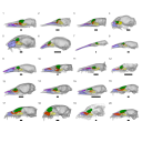
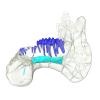
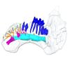
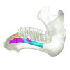
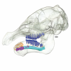
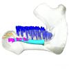
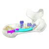
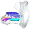
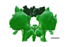
This contribution contains the three-dimensional models of the turbinal complex of 10 myrmecophagous and 10 non-myrmecophagous placental species. These specimens were analyzed and discussed in: Wright et. al (2024), Sniffing out morphological convergence in the turbinal complex of myrmecophagous placentals. https://doi.org/10.1002/ar.25603
Priodontes maximus NHMUK 732-a View specimen

|
M3#1536Turbinals of Priodontes maximus Type: "3D_surfaces"doi: 10.18563/m3.sf.1536 state:published |
Download 3D surface file |
Dasypus pilosus NHMUK 94-10-1-13 View specimen
|
M3#1537Turbinals of Dasypus pilosus Type: "3D_surfaces"doi: 10.18563/m3.sf.1537 state:published |
Download 3D surface file |
Dasypus novemcinctus AMNH 263287 View specimen

|
M3#1538Turbinals of Dasypus novemcinctus Type: "3D_surfaces"doi: 10.18563/m3.sf.1538 state:published |
Download 3D surface file |
Bradypus tridactylus UM 789N View specimen
|
M3#1539Turbinals of Bradypus tridactylus Type: "3D_surfaces"doi: 10.18563/m3.sf.1539 state:published |
Download 3D surface file |
Choloepus didactylus UM 767V View specimen

|
M3#1540Turbinals of Choloepus didactylus Type: "3D_surfaces"doi: 10.18563/m3.sf.1540 state:published |
Download 3D surface file |
Cyclopes didactylus NHMUK 88-8-8-14 View specimen
|
M3#1541Turbinals of Cyclopes didactylus Type: "3D_surfaces"doi: 10.18563/m3.sf.1541 state:published |
Download 3D surface file |
Myrmecophaga tridactyla UM 065V View specimen
|
M3#1542Turbinals of Myrmecophaga tridactyla Type: "3D_surfaces"doi: 10.18563/m3.sf.1542 state:published |
Download 3D surface file |
Tamandua tetradactyla NHMUK 3-7-7-135 View specimen
|
M3#1543Turbinals of Tamandua tetradactyla Type: "3D_surfaces"doi: 10.18563/m3.sf.1543 state:published |
Download 3D surface file |
Tamandua mexicana NHMUK 79-1-6-1 View specimen
|
M3#1544Turbinals of Tamandua mexicana Type: "3D_surfaces"doi: 10.18563/m3.sf.1544 state:published |
Download 3D surface file |
Orycteropus afer NHMUK 2-9-9-58 View specimen
|
M3#1545Turbinals of Orycteropus afer Type: "3D_surfaces"doi: 10.18563/m3.sf.1545 state:published |
Download 3D surface file |
Tenrec eucaudatus UM N439 View specimen
|
M3#1546Turbinals of Tenrec eucaudatus Type: "3D_surfaces"doi: 10.18563/m3.sf.1546 state:published |
Download 3D surface file |
Elephantulus rozeti UM N227 View specimen
|
M3#1547Turbinals of Elephantulus rozeti Type: "3D_surfaces"doi: 10.18563/m3.sf.1547 state:published |
Download 3D surface file |
Phataginus tetradactyla NHMUK 1-11-21-35 View specimen
|
M3#1548Turbinals of Phataginus tetradactyla Type: "3D_surfaces"doi: 10.18563/m3.sf.1548 state:published |
Download 3D surface file |
Smutsia gigantea KMMA 25479 View specimen
|
M3#1549Turbinals of Smutsia gigantea Type: "3D_surfaces"doi: 10.18563/m3.sf.1549 state:published |
Download 3D surface file |
Manis culionensis MNHN ZM-MO 1884-1822 View specimen

|
M3#1550Turbinals of Manis culionensis Type: "3D_surfaces"doi: 10.18563/m3.sf.1550 state:published |
Download 3D surface file |
Vulpes vulpes UM N140 View specimen
|
M3#1551Turbinals of Vulpes vulpes Type: "3D_surfaces"doi: 10.18563/m3.sf.1551 state:published |
Download 3D surface file |
Alopex lagopus UM N110 View specimen
|
M3#1552Turbinals of Alopex lagopus Type: "3D_surfaces"doi: 10.18563/m3.sf.1552 state:published |
Download 3D surface file |
Felis silvestris UM N149 View specimen
|
M3#1553Turbinals of Felis sylvestris Type: "3D_surfaces"doi: 10.18563/m3.sf.1553 state:published |
Download 3D surface file |
Hyaena hyaena UM N109 View specimen
|
M3#1554Turbinals of Hyaena hyaena Type: "3D_surfaces"doi: 10.18563/m3.sf.1554 state:published |
Download 3D surface file |
Proteles cristata NHMUK 4-3-1-58 View specimen
|
M3#1555Turbinals of Proteles cristata Type: "3D_surfaces"doi: 10.18563/m3.sf.1555 state:published |
Download 3D surface file |


