Explodable 3D Dog Skull for Veterinary Education
3D models of Ocnotherium skull
3D models of Kalakocetus, the earliest Cetacea
3D GM dataset of bird skeletal variation
Skeletal embryonic development in the catshark
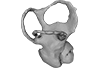
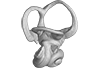
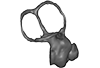
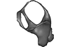
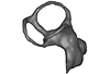
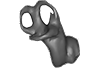
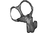
Bony connexions of the petrosal bone of extant hippos
bony labyrinth (14) , inner ear (11) , geometric morphometrics (10) , CT-scan (10) , Eocene (10) , Micro-CT (9) , Miocene (8)
Lionel Hautier (24) , Maëva Judith Orliac (23) , Laurent Marivaux (18) , Renaud Lebrun (14) , Rodolphe Tabuce (14) , Pierre-Olivier Antoine (13) , Bastien Mennecart (13)
|
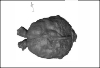
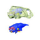
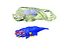
3D data and models related to the publication: An updated description of the osteology of the pancake tortoise Malacochersus tornieri (Testudines: Testudinidae) with special focus on intraspecific variation.Anna-Katharina Mautner
Published online: 25/01/2017 |
|
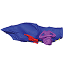
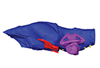
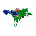
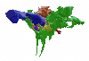
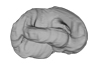
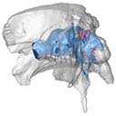
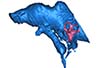
M3#129Virtual brain and inner ear endocast of Malacochersus tornieri (ZM 100.102; Zoological Museum of The University of Zurich). This virtual model is accompanied by the 3D dataset. Blue, endocranium; red, blood vessels; purple, semicircular canals; yellow, cranial nerves. Type: "3D_surfaces"doi: 10.18563/m3.sf.129 state:published |
Download 3D surface file |
|
M3#1303D dataset of skull of Malacochersus tornieri (ZM 100.102) Type: "3D_CT"doi: 10.18563/m3.sf.130 state:published |
Download CT data |
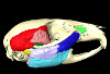
This contribution contains the 3D model(s) described and figured in the following publication: Da Cunha, L., Fabre, P.-H. & Hautier, L. (2024) Springhares, flying and flightless scaly-tailed squirrels (Anomaluromorpha, Rodentia) are the squirrely mouse: comparative anatomy of the masticatory musculature and its implications on the evolution of hystricomorphy in rodents. Journal of Anatomy, 244, 900–928.
Anomalurus derbianus 21804 View specimen
|
M3#1493Masticatory apparatus of Anomalurus Type: "3D_surfaces"doi: 10.18563/m3.sf.1493 state:published |
Download 3D surface file |
Idiurus macrotis 29335 View specimen
|
M3#1492Masticatory apparatus of Idiurus Type: "3D_surfaces"doi: 10.18563/m3.sf.1492 state:published |
Download 3D surface file |
Zenkerella insignis 5.5.23.27 View specimen
|
M3#1490Masticatory apparatus of Zenkerella Type: "3D_surfaces"doi: 10.18563/m3.sf.1490 state:published |
Download 3D surface file |
Pedetes capensis NA View specimen
|
M3#1491Masticatory apparatus of Pedetes Type: "3D_surfaces"doi: 10.18563/m3.sf.1491 state:published |
Download 3D surface file |
The present 3D Dataset contains the 3D models analyzed in Pochat-Cottilloux Y., Rinder N., Perrichon G., Adrien J., Amiot R., Hua S. & Martin J. E. (2023). The neuroanatomy and pneumaticity of Hamadasuchus from the Cretaceous of Morocco and its significance for the paleoecology of Peirosauridae and other altirostral crocodylomorphs. Journal of Anatomy, https://doi.org/10.1111/joa.13887
Hamadasuchus sp. UCBL-FSL 532408 View specimen
|
M3#10943D volume reconstruction of the braincase osteology Type: "3D_surfaces"doi: 10.18563/m3.sf.1094 state:published |
Download 3D surface file |
|
M3#10963D volume reconstruction of the endocast Type: "3D_surfaces"doi: 10.18563/m3.sf.1096 state:published |
Download 3D surface file |
|
M3#10973D volume reconstruction of the labyrinths Type: "3D_surfaces"doi: 10.18563/m3.sf.1097 state:published |
Download 3D surface file |
|
M3#10983D volume reconstruction of the pneumatic cavities Type: "3D_surfaces"doi: 10.18563/m3.sf.1098 state:published |
Download 3D surface file |
The present 3D Dataset contains the 3D models illustrated and described in the chapter “Paleoneurology of Artiodactyla, an overview of the evolution of the artiodactyl brain” (Orliac et al. 2022) published in "Paleoneurology of amniotes: new directions in the study of fossil endocasts", edited by Dozo, Paulina-Carabajal, Macrini and Walsh.
Homacodon vagans AMNH 12695 View specimen
|
M3#1063Endocranial cast Type: "3D_surfaces"doi: 10.18563/m3.sf.1063 state:published |
Download 3D surface file |
Helohyus sp. AMNH 13079 View specimen
|
M3#1064Endocranial cast Type: "3D_surfaces"doi: 10.18563/m3.sf.1064 state:published |
Download 3D surface file |
Leptauchenia sp. AMNH 45508 View specimen
|
M3#1065endocranial cast Type: "3D_surfaces"doi: 10.18563/m3.sf.1065 state:published |
Download 3D surface file |
Agriochoerus sp. AMNH 95330 View specimen
|
M3#1067endocranial cast Type: "3D_surfaces"doi: 10.18563/m3.sf.1067 state:published |
Download 3D surface file |
Mouillacitherium elegans UM ACQ 6625 View specimen
|
M3#1068endocranial cast Type: "3D_surfaces"doi: 10.18563/m3.sf.1068 state:published |
Download 3D surface file |
Caenomeryx filholi UM PDS 2570 View specimen
|
M3#1069endocranial cast Type: "3D_surfaces"doi: 10.18563/m3.sf.1069 state:published |
Download 3D surface file |
Dichobune leporina MNHN.F.QU16586 View specimen
|
M3#1070endocranial cast Type: "3D_surfaces"doi: 10.18563/m3.sf.1070 state:published |
Download 3D surface file |
Anoplotherium sp. not numbered View specimen
|
M3#1071endocranial cast Type: "3D_surfaces"doi: 10.18563/m3.sf.1071 state:published |
Download 3D surface file |
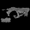
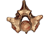
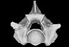
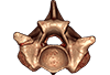
This contribution contains the 3D models described and figured in the following publication: Georgalis, G.L., G. Guinot, K.E. Kassegne, Y.Z. Amoudji, A.K.C. Johnson, H. Cappetta and L. Hautier. 2021. An assemblage of giant aquatic snakes (Serpentes, Palaeophiidae) from the Eocene of Togo. Swiss Journal of Palaeontology 140, https://doi.org/10.1186/s13358-021-00236-w
Palaeophis africanus UM KPO 21 View specimen
|
M3#821Trunk vertebra UM KPO 21 of Palaeophis africanus Type: "3D_surfaces"doi: 10.18563/m3.sf.821 state:published |
Download 3D surface file |
Palaeophis africanus UM KPO 22 View specimen
|
M3#822Trunk vertebra UM KPO 22 of Palaeophis africanus from the Eocene of Togo Type: "3D_surfaces"doi: 10.18563/m3.sf.822 state:published |
Download 3D surface file |
Palaeophis africanus UM KPO 23 View specimen
|
M3#823Trunk vertebra UM KPO 23 of Palaeophis africanus Type: "3D_surfaces"doi: 10.18563/m3.sf.823 state:published |
Download 3D surface file |
Palaeophis africanus UM KPO 24 View specimen
|
M3#824Trunk vertebra UM KPO 24 of Palaeophis africanus Type: "3D_surfaces"doi: 10.18563/m3.sf.824 state:published |
Download 3D surface file |
Palaeophis africanus UM KPO 25 View specimen
|
M3#825Trunk vertebra UM KPO 25 of Palaeophis africanus Type: "3D_surfaces"doi: 10.18563/m3.sf.825 state:published |
Download 3D surface file |
Palaeophis africanus UM KPO 26 View specimen
|
M3#826Trunk vertebra UM KPO 26 of Palaeophis africanus Type: "3D_surfaces"doi: 10.18563/m3.sf.826 state:published |
Download 3D surface file |
Palaeophis africanus UM KPO 27 View specimen
|
M3#827Trunk vertebra UM KPO 27 of Palaeophis africanus Type: "3D_surfaces"doi: 10.18563/m3.sf.827 state:published |
Download 3D surface file |
Palaeophis africanus UM KPO 28 View specimen
|
M3#828Trunk vertebra UM KPO 28 of Palaeophis africanus Type: "3D_surfaces"doi: 10.18563/m3.sf.828 state:published |
Download 3D surface file |
Palaeophis africanus UM KPO 29 View specimen
|
M3#829Trunk vertebra UM KPO 29 of Palaeophis africanus Type: "3D_surfaces"doi: 10.18563/m3.sf.829 state:published |
Download 3D surface file |
Palaeophis africanus UM KPO 30 View specimen
|
M3#830Trunk vertebra UM KPO 30 of Palaeophis africanus Type: "3D_surfaces"doi: 10.18563/m3.sf.830 state:published |
Download 3D surface file |
Palaeophis africanus UM KPO 31 View specimen
|
M3#831Trunk vertebra UM KPO 28 of Palaeophis africanus Type: "3D_surfaces"doi: 10.18563/m3.sf.831 state:published |
Download 3D surface file |
Palaeophis africanus UM KPO 32 View specimen
|
M3#832Trunk vertebra UM KPO 32 of Palaeophis africanus Type: "3D_surfaces"doi: 10.18563/m3.sf.832 state:published |
Download 3D surface file |
Palaeophis africanus UM KPO 33 View specimen
|
M3#833Trunk vertebra UM KPO 33 of Palaeophis africanus Type: "3D_surfaces"doi: 10.18563/m3.sf.833 state:published |
Download 3D surface file |
Palaeophis africanus UM KPO 34 View specimen
|
M3#839Trunk vertebra UM KPO 34 of Palaeophis africanus Type: "3D_surfaces"doi: 10.18563/m3.sf.839 state:published |
Download 3D surface file |
Palaeophis africanus UM KPO 35 View specimen
|
M3#840Trunk vertebra UM KPO 35 of Palaeophis africanus Type: "3D_surfaces"doi: 10.18563/m3.sf.840 state:published |
Download 3D surface file |
Palaeophis africanus UM KPO 36 View specimen
|
M3#841Trunk vertebra UM KPO 36 of Palaeophis africanus Type: "3D_surfaces"doi: 10.18563/m3.sf.841 state:published |
Download 3D surface file |
Palaeophis africanus UM KPO 37 View specimen
|
M3#842Trunk vertebra UM KPO 37 of Palaeophis africanus Type: "3D_surfaces"doi: 10.18563/m3.sf.842 state:published |
Download 3D surface file |
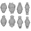
The present 3D Dataset contains 26 3D models analyzed in the study: On the “cartilaginous rider” in the endocasts of turtle brain cavities, published by the authors in the journal Vertebrate Zoology.
Annemys sp. IVPP-V-18106 View specimen
|
M3#7723D surface(s) file to specimen IVPP-V-18106 Type: "3D_surfaces"doi: 10.18563/m3.sf.772 state:published |
Download 3D surface file |
Apalone spinifera FMNH 22178 View specimen

|
M3#7733D surface(s) file to specimen FMNH 22178 Type: "3D_surfaces"doi: 10.18563/m3.sf.773 state:published |
Download 3D surface file |
Caretta caretta NHMUK1940.3.15.1 View specimen
|
M3#7863D surface(s) file to specimen NHMUK1940.3.15.1 Type: "3D_surfaces"doi: 10.18563/m3.sf.786 state:published |
Download 3D surface file |
Chelodina reimanni ZMB 49659 View specimen

|
M3#7743D surface(s) file to specimen ZMB 49659 Type: "3D_surfaces"doi: 10.18563/m3.sf.774 state:published |
Download 3D surface file |
Chelonia mydas ZMB-37416MS View specimen
|
M3#7753D surface(s) file to specimen ZMB-37416MS Type: "3D_surfaces"doi: 10.18563/m3.sf.775 state:published |
Download 3D surface file |
Cuora amboinensis NHMUK69.42.145_4 View specimen
|
M3#7763D surface(s) file to specimen NHMUK69.42.145_4 Type: "3D_surfaces"doi: 10.18563/m3.sf.776 state:published |
Download 3D surface file |
Emydura subglobosa IW92 View specimen
|
M3#7773D surface(s) file to specimen IW92 Type: "3D_surfaces"doi: 10.18563/m3.sf.777 state:published |
Download 3D surface file |
Eubaena cephalica DMNH 96004 View specimen
|
M3#7783D surface(s) file to specimen DMNH 96004 Type: "3D_surfaces"doi: 10.18563/m3.sf.778 state:published |
Download 3D surface file |
Gopherus berlandieri AMNH-73816 View specimen
|
M3#7793D surface(s) file to specimen AMNH-73816 Type: "3D_surfaces"doi: 10.18563/m3.sf.779 state:published |
Download 3D surface file |
Kinixys belliana AMNH-10028 View specimen
|
M3#7803D surface(s) file to specimen AMNH-10028 Type: "3D_surfaces"doi: 10.18563/m3.sf.780 state:published |
Download 3D surface file |
Macrochelys temminckii GPIT-PV-79430 View specimen
|
M3#7813D surface(s) file to specimen GPIT-PV-79430 Type: "3D_surfaces"doi: 10.18563/m3.sf.781 state:published |
Download 3D surface file |
Malacochersus tornieri SMF-58702 View specimen
|
M3#7873D surface(s) file to specimen SMF-58702 Type: "3D_surfaces"doi: 10.18563/m3.sf.787 state:published |
Download 3D surface file |
Naomichelys speciosa FMNH-PR-273 View specimen
|
M3#7823D surface(s) file to specimen FMNH-PR-273 Type: "3D_surfaces"doi: 10.18563/m3.sf.782 state:published |
Download 3D surface file |
Pelodiscus sinensis IW576-2 View specimen
|
M3#7833D surface(s) file to specimen IW576-2 Type: "3D_surfaces"doi: 10.18563/m3.sf.783 state:published |
Download 3D surface file |
Platysternon megacephalum SMF-69684 View specimen
|
M3#7843D surface(s) file to specimen SMF-69684 Type: "3D_surfaces"doi: 10.18563/m3.sf.784 state:published |
Download 3D surface file |
Podocnemis unifilis SMF-55470 View specimen
|
M3#7853D surface(s) file to specimen SMF-55470 Type: "3D_surfaces"doi: 10.18563/m3.sf.785 state:published |
Download 3D surface file |
Proganochelys quenstedtii MB 1910.45.2 View specimen
|
M3#7883D surface(s) file to specimen MB 1910.45.2 Type: "3D_surfaces"doi: 10.18563/m3.sf.788 state:published |
Download 3D surface file |
Proganochelys quenstedtii SMNS 16980 View specimen
|
M3#7893D surface(s) file to specimen SMNS 16980 Type: "3D_surfaces"doi: 10.18563/m3.sf.789 state:published |
Download 3D surface file |
Rhinochelys pulchriceps CAMSM_B55775 View specimen
|
M3#7903D surface(s) file to specimen CAMSM_B55775 Type: "3D_surfaces"doi: 10.18563/m3.sf.790 state:published |
Download 3D surface file |
Rhinoclemmys funereal YPM12174 View specimen
|
M3#7913D surface(s) file to specimen YPM12174 Type: "3D_surfaces"doi: 10.18563/m3.sf.791 state:published |
Download 3D surface file |
Sandownia harrisi MIWG3480 View specimen
|
M3#7923D surface(s) file to specimen MIWG3480 Type: "3D_surfaces"doi: 10.18563/m3.sf.792 state:published |
Download 3D surface file |
Testudo graeca YPM14342 View specimen
|
M3#7933D surface(s) file to specimen YPM14342 Type: "3D_surfaces"doi: 10.18563/m3.sf.793 state:published |
Download 3D surface file |
Testudo hermanni AMNH134518 View specimen
|
M3#7943D surface(s) file to specimen AMNH134518 Type: "3D_surfaces"doi: 10.18563/m3.sf.794 state:published |
Download 3D surface file |
Trachemys scripta NN View specimen
|
M3#7953D surface(s) file to specimen Trachemys scripta Type: "3D_surfaces"doi: 10.18563/m3.sf.795 state:published |
Download 3D surface file |
Xinjiangchelys radiplicatoides IVPP V9539 View specimen
|
M3#7963D surface(s) file to specimen IVPP V9539 Type: "3D_surfaces"doi: 10.18563/m3.sf.796 state:published |
Download 3D surface file |
Chelydra serpentina UFR VP1 View specimen
|
M3#801Brain endocast Type: "3D_surfaces"doi: 10.18563/m3.sf.801 state:published |
Download 3D surface file |
The present 3D Dataset contains the 3D models of the holotype and the paratypes of the new species Siphonodella leiosa described and analyzed in the following publication: L. Souquet, C. Corradini, C. Girard: Siphonodella leiosa (Conodonta), a new unornamented species from the Tournaisian (lower Carboniferous) of Puech de la Suque (Montagne Noire, France). Geobios, https://doi.org/10.1016/j.geobios.2020.06.004.
Siphonodella leiosa UM PSQ 1 View specimen
|
M3#525Siphonodella leiosa, paratype, dextral P1 element Type: "3D_surfaces"doi: 10.18563/m3.sf.525 state:published |
Download 3D surface file |
Siphonodella leiosa UM PSQ 2 View specimen
|
M3#526Siphonodella leiosa, holotype, dextral P1 element Type: "3D_surfaces"doi: 10.18563/m3.sf.526 state:published |
Download 3D surface file |
Siphonodella leiosa UM PSQ 3 View specimen
|
M3#527Siphonodella leiosa, paratype, dextral P1 element Type: "3D_surfaces"doi: 10.18563/m3.sf.527 state:published |
Download 3D surface file |
Siphonodella leiosa UM PSQ 4 View specimen

|
M3#528Siphonodella leiosa, paratype, dextral P1 element Type: "3D_surfaces"doi: 10.18563/m3.sf.528 state:published |
Download 3D surface file |
Siphonodella leiosa UM PSQ 5 View specimen
|
M3#529Siphonodella leiosa, paratype, sinistral P1 element Type: "3D_surfaces"doi: 10.18563/m3.sf.529 state:published |
Download 3D surface file |
Siphonodella leiosa UM PSQ 6 View specimen
|
M3#530Siphonodella leiosa, paratype, dextral P1 element Type: "3D_surfaces"doi: 10.18563/m3.sf.530 state:published |
Download 3D surface file |
Siphonodella leiosa UM PSQ 7 View specimen
|
M3#531Siphonodella leiosa, paratype, dextral P1 element Type: "3D_surfaces"doi: 10.18563/m3.sf.531 state:published |
Download 3D surface file |
Siphonodella leiosa UM PSQ 8 View specimen
|
M3#532Siphonodella leiosa, paratype, sinistral P1 element Type: "3D_surfaces"doi: 10.18563/m3.sf.532 state:published |
Download 3D surface file |
Siphonodella leiosa UM PSQ 9 View specimen
|
M3#533Siphonodella leiosa, paratype, dextral P1 element Type: "3D_surfaces"doi: 10.18563/m3.sf.533 state:published |
Download 3D surface file |
The present 3D Dataset contains the 3D surface model and the µCT scan analyzed in the following publication: R. Tabuce, R. Sarr, S. Adnet, R. Lebrun, F. Lihoreau, J. E. Martin, B. Sambou, M. Thiam, and L. Hautier: Filling a gap in the proboscidean fossil record: a new genus from the Lutetian of Senegal. Journal of Paleontology, in press, doi: 10.1017/jpa.2019.98
Saloumia gorodiskii MNHN.F.MCA 1 View specimen
|
M3#500Tooth 3D model of Saloumia gorodiskii Type: "3D_surfaces"doi: 10.18563/m3.sf500 state:published |
Download 3D surface file |
|
M3#501µCT scan of Saloumia gorodiskii Type: "3D_CT"doi: 10.18563/m3.sf501 state:published |
Download CT data |
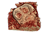
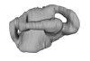
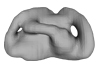
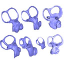
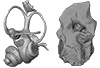
The present 3D Dataset contains the 3D models analyzed in the publication ‘Ontogenetic development of the otic region in the new model organism, Leucoraja erinacea (Chondrichthyes; Rajidae)’, https://doi.org/10.1017/S1755691018000993
Leucoraja erinacea 2018.9.26.1 View specimen
|
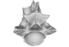
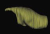
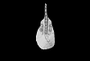
M3#3673D model of the right skeletal labyrinth of the adult specimen of Leucoraja erincea. T Type: "3D_surfaces"doi: 10.18563/m3.sf.367 state:published |
Download 3D surface file |
Leucoraja erinacea 2018.9.25.2 View specimen
|
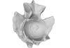
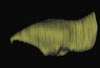
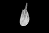
M3#3683D model of the right skeletal labyrinth of the stage 34 specimen of Leucoraja erincea. Type: "3D_surfaces"doi: 10.18563/m3.sf.368 state:published |
Download 3D surface file |
Leucoraja erinacea 2018.9.25.3 View specimen
|
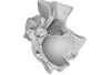
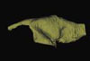
M3#3693D model of the right skeletal labyrinth of the stage 32 specimen of Leucoraja erinacea. Type: "3D_surfaces"doi: 10.18563/m3.sf.369 state:published |
Download 3D surface file |
|
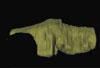
M3#3723D model of the right membranous system of stage 32 of Leucoraja erincea. Type: "3D_surfaces"doi: 10.18563/m3.sf.372 state:published |
Download 3D surface file |
Leucoraja erinacea 2018.9.25.4 View specimen
|
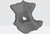
M3#3703D model of the right skeletal labyrinth of the stage 31 specimen of Leucoraja erinacea. Type: "3D_surfaces"doi: 10.18563/m3.sf.370 state:published |
Download 3D surface file |
Leucoraja erinacea 2018.9.26.5 View specimen

|
M3#3763D model of the right skeletal labyrinth of the stage 29 specimen of Leucoraja erinacea. Type: "3D_surfaces"doi: 10.18563/m3.sf.376 state:published |
Download 3D surface file |
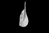
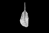
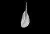
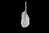
Here, the semicircular canals of the most aquatic seal, the rare Antarctic Ross Seal (Ommatophoca rossii), are presented for the first time, along with representatives of every species in the Lobodontini: the leopard seal (Hydrurga leptonyx), Weddell seal (Leptonychotes weddellii), and crabeater seal (Lobodon carcinophagus). Because encounters with wild Ross seal are rare, and few specimens are available in collections worldwide, this dataset increases accessibility to a rare species. For further comparison, we present the bony labyrinths of other carnivorans, the elephant seal (Mirounga leonina), harbor seal (Phoca vitulina), walrus (Odobenus rosmarus), South American sea lion (Otaria byronia).
Odobenus rosmarus MVZ 125566 View specimen
|
M3#173Surface of the semicircular canals and cochlea of the walrus, Odobenus rosmarus Type: "3D_surfaces"doi: 10.18563/m3.sf.173 state:published |
Download 3D surface file |
Phoca vitulina UZNH 17973 View specimen
|
M3#174Endocast surface of the semicircular canals and cochlea of the harbor seal, Phoca vitulina. Type: "3D_surfaces"doi: 10.18563/m3.sf.174 state:published |
Download 3D surface file |
Hydrurga leptonyx MLP 14.IV.48.11 View specimen
|
M3#285Endocast surface of the semicircular canals and cochlea of the leopard seal, Hydrurga leptonyx. Type: "3D_surfaces"doi: 10.18563/m3.sf.285 state:published |
Download 3D surface file |
Leptonychotes weddellii IAA 02-13 View specimen
|
M3#288Endocast surface of the semicircular canals and cochlea of the Weddell seal Leptonychotes weddellii. Type: "3D_surfaces"doi: 10.18563/m3.sf.288 state:published |
Download 3D surface file |
Lobodon carcinophagus IAA 530 View specimen
|
M3#286Endocast surface of the semicircular canals and cochlea of the crabeater seal, Lobodon carcinophagus. Type: "3D_surfaces"doi: 10.18563/m3.sf.286 state:published |
Download 3D surface file |
Ommatophoca rossii MACN 48259 View specimen
|
M3#176Endocast surface of the semicircular canals and cochlea of the Ross seal Ommatophoca rossii. Type: "3D_surfaces"doi: 10.18563/m3.sf.176 state:published |
Download 3D surface file |
Mirounga leonina IAA 03-5 View specimen
|
M3#287Right endocast surface of the semicircular canals and cochlea of the elephant seal, Mirounga leonina. Type: "3D_surfaces"doi: 10.18563/m3.sf.287 state:published |
Download 3D surface file |
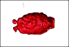
This contribution contains the 3D models described and figured in the following publication: Paulina-Carabajal A and Calvo JO 2021. Re-description of the braincase of the rebbachisaurid sauropod Limaysaurus tessonei and novel endocranial information based on CT scans. Anais da Academia Brasileira de Ciências 93(Suppl. 2): e20200762 https://doi.org/10.1590/0001-3765202120200762
Limaysaurus tessonei MUCPv-205 View specimen
|
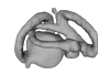
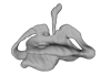
M3#700Renderings of the virtually isolate braincase, brain, and right inner ear. Type: "3D_surfaces"doi: 10.18563/m3.sf.700 state:published |
Download 3D surface file |
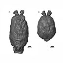
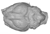
The present 3D Dataset contains the 3D models of the brain endocast analyzed in “Virtual brain endocast of Antifer (Mammalia: Cervidae), an extinct large cervid from South America”.
Antifer ensenadensis U-4922 View specimen
|
M3#550Brain endocast Type: "3D_surfaces"doi: 10.18563/m3.sf.550 state:published |
Download 3D surface file |
Antifer ensenadensis MCN-PV 943 View specimen
|
M3#551Brain endocast Type: "3D_surfaces"doi: 10.18563/m3.sf.551 state:published |
Download 3D surface file |
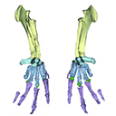
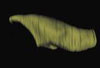
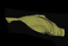
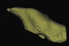
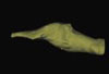
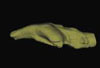
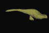
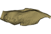
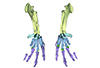
The present 3D Dataset contains the 3D model analyzed in Vautrin et al. (2019), Palaeontology, From limb to fin: an Eocene protocetid forelimb from Senegal sheds new light on the early locomotor evolution of early cetaceans.
?Carolinacetus indet. SNTB 2011-01 View specimen
|
M3#3983D model of an articulated forelimb of a Carolinacetus-like protocetid from Senegal Type: "3D_surfaces"doi: 10.18563/m3.sf.398 state:published |
Download 3D surface file |
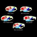
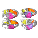
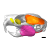
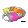
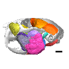
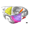
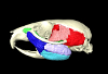
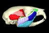
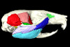
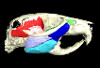
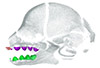
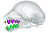
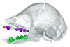
The present 3D Dataset contains the 3D models analyzed in the article entitled "One skull to rule them all? Descriptive and comparative anatomy of the masticatory apparatus in five mice species based on traditional and digital dissections" (Ginot et al. 2018, Journal of Morphology, https://doi.org/10.1002/jmor.20845).
Mus cervicolor R7314 View specimen
|
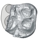
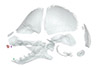
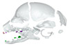
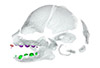
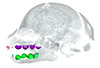
M3#343.ply surfaces of the skull and masticatory muscles of Mus cervicolor. Created with MorphoDig, .pos and .ntw files also included. Scans were obtained thanks to the Institut des Sciences de l'Evolution de Montpellier MRI platform. Type: "3D_surfaces"doi: 10.18563/m3.sf.343 state:published |
Download 3D surface file |
Mus caroli R7264 View specimen
|
M3#344.ply surfaces of the skull and masticatory muscles of Mus caroli. Created with MorphoDig, .pos and .ntw files also included. Scans were obtained thanks to the Institut des Sciences de l'Evolution de Montpellier MRI platform. Type: "3D_surfaces"doi: 10.18563/m3.sf.344 state:published |
Download 3D surface file |
Mus fragilicauda R7260 View specimen
|
M3#345.ply surfaces of the skull and masticatory muscles of Mus fragilicauda. Created with MorphoDig, .pos and .ntw files also included. Scans were obtained thanks to the Institut des Sciences de l'Evolution de Montpellier MRI platform. Type: "3D_surfaces"doi: 10.18563/m3.sf.345 state:published |
Download 3D surface file |
Mus pahari R7226 View specimen
|
M3#346.ply surfaces of the skull and masticatory muscles of Mus pahari. Created with MorphoDig, .pos and .ntw files also included. Scans were obtained thanks to the Institut des Sciences de l'Evolution de Montpellier MRI platform. Type: "3D_surfaces"doi: 10.18563/m3.sf.346 state:published |
Download 3D surface file |
Mus minutoides minutoides-1 View specimen
|
M3#347.ply surfaces of the skull and masticatory muscles of Mus minutoides. Created with MorphoDig, .pos and .ntw files also included. Scans were obtained thanks to the Institut des Sciences de l'Evolution de Montpellier MRI platform. Type: "3D_surfaces"doi: 10.18563/m3.sf.347 state:published |
Download 3D surface file |
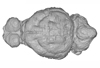
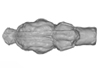
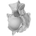
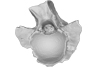
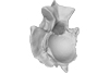
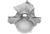
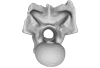
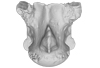
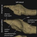
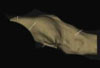
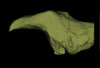
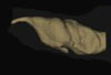
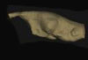
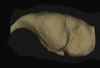
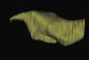
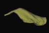
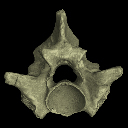
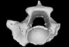
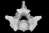
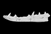
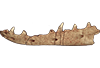
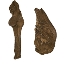
This contribution contains the 3D models described and figured in the following publication: Mennecart B., de Perthuis Ad., Rössner G.E., Guzmán J.A., de Perthuis Au., Costeur L. The first French tragulid skull (Mammalia, Ruminantia, Tragulidae) and associated tragulid remains from the Middle Miocene of Contres (Loir-et-Cher, France). Comptes Rendus Palévol. https://doi.org/10.1016/j.crpv.2017.08.004
Dorcatherium crassum NMB Fa.213.abg View specimen
|
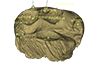
M3#181The 3D surface files of the specimen NMB Fa.213 are the reconstructions of the main skull fragments, the right petrosal bone, and the left bony labyrinth. Type: "3D_surfaces"doi: 10.18563/m3.sf.181 state:published |
Download 3D surface file |
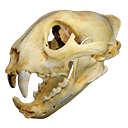
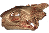
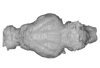
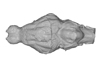
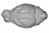
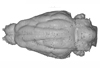
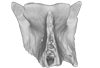
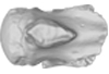
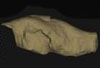
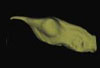
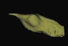
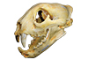
The present 3D Dataset contains the 3D model of a skull analyzed in “A Puma concolor (Carnivora: Felidae) in the Middle-Late Holocene landscapes of the Brazilian Northeast (Bahia): submerged cave deposits and stable isotopes”. The 3D model was generated by photogrammetry.
Puma concolor MN 57461 View specimen
|
M3#843Cranium Type: "3D_surfaces"doi: 10.18563/m3.sf.843 state:published |
Download 3D surface file |
The present 3D Dataset contains the 3D models analyzed in the following publication: Georgalis, G. L., and T. M. Scheyer. A new species of Palaeopython (Serpentes) and other extinct squamates from the Eocene of Dielsdorf (Zurich, Switzerland). Swiss Journal of Geosciences (in press). https://doi.org/10.1007/s00015-019-00341-6
Palaeopython helveticus PIMUZ A/III 631 View specimen
|
M3#399ZIP file containing .ply of vertebra PIMUZ A/III 631 from Palaeopython helveticus n. sp. Type: "3D_surfaces"doi: 10.18563/m3.sf.399 state:published |
Download 3D surface file |
|
M3#403dataset of snake vertebra PIMUZ A/III 631 Type: "3D_CT"doi: 10.18563/m3.sf.403 state:published |
Download CT data |
Palaeopython helveticus PIMUZ A/III 634 View specimen
|
M3#400ZIP file containing .ply of vertebra PIMUZ A/III 634 from Palaeopython helveticus n. sp. (holotype) Type: "3D_surfaces"doi: 10.18563/m3.sf.400 state:published |
Download 3D surface file |
|
M3#404dataset of snake vertbra PIMUZ A/III 634 (holotype) Type: "3D_CT"doi: 10.18563/m3.sf.404 state:published |
Download CT data |
Palaeopython helveticus PIMUZ A/III 636 View specimen
|
M3#401ZIP file containing .ply of vertebra PIMUZ A/III 636 from Palaeopython helveticus n. sp. Type: "3D_surfaces"doi: 10.18563/m3.sf.401 state:published |
Download 3D surface file |
|
M3#406dataset of snake vertebra PIMUZ A/III 636 Type: "3D_CT"doi: 10.18563/m3.sf.406 state:published |
Download CT data |
Palaeovaranus sp. PIMUZ A/III 234 View specimen
|
M3#402ZIP file containing .ply of dentary PIMUZ A/III 234 of Palaeovaranus sp. Type: "3D_surfaces"doi: 10.18563/m3.sf.402 state:published |
Download 3D surface file |
|
M3#405dataset of dentary of Palaeovaranus sp. (PIMUZ A/III 234) Type: "3D_CT"doi: 10.18563/m3.sf.405 state:published |
Download CT data |
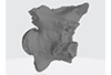
This contribution contains the 3D model described and figured in the following publication: New turtles from the Late Cretaceous of Monte Alto-SP, Brazil, including cranial osteology, neuroanatomy and phylogenetic position of a new taxon. PalZ. https://doi.org/10.1007/s12542-017-0397-x
Yuraramirim montealtensis 04-0008/89 View specimen
|
M3#2783D surfaces related to specimen MPMA 04-0008/89. Type: "3D_surfaces"doi: 10.18563/m3.sf.278 state:published |
Download 3D surface file |
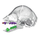
This contribution contains the 3D models described and figured in the following publication: Hautier L., Gomes Rodrigues H., Billet G., Asher R.J., 2016. The hidden teeth of sloths: evolutionary vestiges and the development of a simplified dentition. Scientific Reports. doi: 10.1038/srep27763
Bradypus variegatus ZMB 33812 View specimen
|
M3#110Three-dimensional reconstruction of the teeth, mandibles, maxillary and premaxillary bones Type: "3D_surfaces"doi: 10.18563/m3.sf.110 state:published |
Download 3D surface file |
Bradypus variegatus ZMB 41122 View specimen
|
M3#109Three-dimensional reconstruction of the teeth, mandibles, maxillary and premaxillary bones Type: "3D_surfaces"doi: 10.18563/m3.sf.109 state:published |
Download 3D surface file |
Bradypus variegatus MNHN-ZM-MO-1995-326A View specimen
|
M3#111Three-dimensional reconstruction of the teeth, mandibles, maxillary and premaxillary bones Type: "3D_surfaces"doi: 10.18563/m3.sf.111 state:published |
Download 3D surface file |
Bradypus variegatus MNHN-ZM-MO-1995-326B View specimen
|
M3#112Three-dimensional reconstruction of the teeth, mandibles, maxillary and premaxillary bones Type: "3D_surfaces"doi: 10.18563/m3.sf.112 state:published |
Download 3D surface file |
Bradypus sp. MNHN-ZM-MO-1902-325 View specimen
|
M3#113Three-dimensional reconstruction of the teeth, mandibles, maxillary, and premaxillary bones Type: "3D_surfaces"doi: 10.18563/m3.sf.113 state:published |
Download 3D surface file |
Bradypus sp. MNHN-ZM-MO-1995-327 View specimen
|
M3#114Three-dimensional reconstruction of the teeth, mandibles, maxillary and premaxillary bones Type: "3D_surfaces"doi: 10.18563/m3.sf.114 state:published |
Download 3D surface file |
Choloepus didactylus MNHN-ZM-MO-1882-625 View specimen
|
M3#115Three-dimensional reconstruction of the teeth, mandibles, maxillary and premaxillary bones Type: "3D_surfaces"doi: 10.18563/m3.sf.115 state:published |
Download 3D surface file |
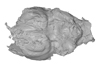
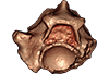
The present 3D Dataset contains the 3D models analyzed in Neogene sloth assemblages (Mammalia, Pilosa) of the Cocinetas Basin (La Guajira, Colombia): implications for the Great American Biotic Interchange. Palaeontology. doi: 10.1111/pala.12244
cf. Nothrotherium indet. MUN STRI 12924 View specimen
|
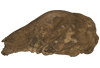
M3#106Fragmentary basicranium with posterior portion of the skull roof. Type: "3D_surfaces"doi: 10.18563/m3.sf.106 state:published |
Download 3D surface file |
indet. indet. MUN STRI 16535 View specimen
|
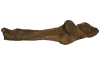
M3#107Complete left ulna of a Scelidotheriinae gen. et sp. indet. Type: "3D_surfaces"doi: 10.18563/m3.sf.107 state:published |
Download 3D surface file |


