Explodable 3D Dog Skull for Veterinary Education
3D models of Ocnotherium skull
3D models of Kalakocetus, the earliest Cetacea
3D GM dataset of bird skeletal variation
Skeletal embryonic development in the catshark
Bony connexions of the petrosal bone of extant hippos
bony labyrinth (14) , inner ear (11) , geometric morphometrics (10) , CT-scan (10) , Eocene (10) , Micro-CT (9) , Miocene (8)
Lionel Hautier (24) , Maëva Judith Orliac (23) , Laurent Marivaux (18) , Renaud Lebrun (14) , Rodolphe Tabuce (14) , Pierre-Olivier Antoine (13) , Bastien Mennecart (13)
|
3D model related to the publication: The endocranial anatomy of the stem turtle Naomichelys speciosa from the Early Cretaceous of North AmericaAriana Paulina-Carabajal
Published online: 10/09/2019 |

|
M3#428FMNH_PR273_1 - Naomichlys speciosa - skull Type: "3D_surfaces"doi: 10.18563/m3.sf.428 state:published |
Download 3D surface file |
This contribution contains the 3D model described and figured in the following publication: Hautier L, Sarr R, Lihoreau F, Tabuce R, Marwan Hameh P. 2014. First record of the family Protocetidae in the Lutetian of Senegal (West Africa). Palaeovertebrata 38(2)-e2
indet. indet. SN103 View specimen

|
M3#5SN103, partial left innominate. Age and occurrence – Taïba Formation, Lutetian of the near Taïba Ndiaye, quarry of the Industries Chimiques du Sénégal (ICS) Type: "3D_surfaces"doi: 10.18563/m3.sf5 state:published |
Download 3D surface file |
This contribution contains the 3D models of postcranial bones (humerus, ulna, innominate, femur, tibia, astragalus, navicular, and metatarsal III) described and figured in the following publication: “Postcranial morphology of the extinct rodent Neoepiblema (Rodentia: Chinchilloidea): insights into the paleobiology of neoepiblemids”.
Neoepiblema acreensis UFAC 3549 View specimen
|
M3#719UFAC 3549, left humerus missing the proximal region. Type: "3D_surfaces"doi: 10.18563/m3.sf.719 state:published |
Download 3D surface file |
Neoepiblema acreensis UFAC 5076 View specimen
|
M3#720UFAC 5076, right humerus missing the proximal region. Type: "3D_surfaces"doi: 10.18563/m3.sf.720 state:published |
Download 3D surface file |
Neoepiblema acreensis UFAC 1939 View specimen
|
M3#721UFAC 1939, right ulna missing the olecranon epiphysis and the distal region. Type: "3D_surfaces"doi: 10.18563/m3.sf.721 state:published |
Download 3D surface file |
Neoepiblema acreensis UFAC 3697 View specimen
|
M3#722UFAC 3697, right innominate bone. Type: "3D_surfaces"doi: 10.18563/m3.sf.722 state:published |
Download 3D surface file |
Neoepiblema acreensis UFAC 2574 View specimen
|
M3#723UFAC 2574, proximal region of a left femur. Type: "3D_surfaces"doi: 10.18563/m3.sf.723 state:published |
Download 3D surface file |
Neoepiblema acreensis UFAC 2937 View specimen
|
M3#724UFAC 2937, right femur with damaged proximal region. Type: "3D_surfaces"doi: 10.18563/m3.sf.724 state:published |
Download 3D surface file |
Neoepiblema acreensis UFAC 2210 View specimen
|
M3#725UFAC 2210, distal region of a right femur. Type: "3D_surfaces"doi: 10.18563/m3.sf.725 state:published |
Download 3D surface file |
Neoepiblema acreensis UFAC 1887 View specimen
|
M3#726UFAC 1887, right tibia Type: "3D_surfaces"doi: 10.18563/m3.sf.726 state:published |
Download 3D surface file |
Neoepiblema acreensis UFAC 1840 View specimen
|
M3#727UFAC 1840, left astragalus. Type: "3D_surfaces"doi: 10.18563/m3.sf.727 state:published |
Download 3D surface file |
Neoepiblema acreensis UFAC 2549 View specimen
|
M3#728UFAC 2549, right astragalus. Type: "3D_surfaces"doi: 10.18563/m3.sf.728 state:published |
Download 3D surface file |
Neoepiblema acreensis UFAC 3672 View specimen
|
M3#729UFAC 3672, right navicular. Type: "3D_surfaces"doi: 10.18563/m3.sf.729 state:published |
Download 3D surface file |
Neoepiblema acreensis UFAC 2116 View specimen
|
M3#730UFAC 2116, left metatarsal III. Type: "3D_surfaces"doi: 10.18563/m3.sf.730 state:published |
Download 3D surface file |
Neoepiblema horridula UFAC 3260 View specimen
|
M3#731UFAC 3260, fragmented left innominate. Type: "3D_surfaces"doi: 10.18563/m3.sf.731 state:published |
Download 3D surface file |
Neoepiblema horridula UFAC 2620 View specimen
|
M3#732UFAC 2620, distal region of a right femur. Type: "3D_surfaces"doi: 10.18563/m3.sf.732 state:published |
Download 3D surface file |
Neoepiblema horridula UFAC 2737 View specimen
|
M3#733UFAC 2737, proximal region of right femur. Type: "3D_surfaces"doi: 10.18563/m3.sf.733 state:published |
Download 3D surface file |
Neoepiblema horridula UFAC 3202 View specimen
|
M3#734UFAC 3202, right tibia, missing the proximalmost and distal portions. Type: "3D_surfaces"doi: 10.18563/m3.sf.734 state:published |
Download 3D surface file |
Neoepiblema horridula UFAC 3212 View specimen
|
M3#735UFAC 3212, left astragalus. Type: "3D_surfaces"doi: 10.18563/m3.sf.735 state:published |
Download 3D surface file |
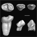
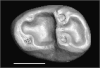
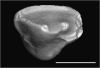
This contribution contains the 3D models of the fossil teeth of two chinchilloid caviomorph rodents (Borikenomys praecursor and Chinchilloidea gen. et sp. indet.) discovered from lower Oligocene deposits of Puerto Rico, San Sebastian Formation (locality LACM Loc. 8060). These fossils were described and figured in the following publication: Marivaux et al. (2020), Early Oligocene chinchilloid caviomorphs from Puerto Rico and the initial rodent colonization of the West Indies. Proceedings of the Royal Society B. http://dx.doi.org/10.1098/rspb.2019.2806
Borikenomys praecursor LACM 162447 View specimen
|
M3#638Right lower m3. This isolated tooth was scanned with a resolution of 6 µm using a μ-CT-scanning station EasyTom 150 / Rx Solutions (Montpellier RIO Imaging, ISE-M, Montpellier, France). AVIZO 7.1 (Visualization Sciences Group) software was used for visualization, segmentation, and 3D rendering. The specimen was prepared within a “labelfield” module of AVIZO, using the segmentation threshold selection tool. Type: "3D_surfaces"doi: 10.18563/m3.sf.638 state:published |
Download 3D surface file |
Borikenomys praecursor LACM 162446 View specimen
|
M3#639Fragment of lower molar (most of the mesial part). This isolated broken tooth was scanned with a resolution of 6 µm using a μ-CT-scanning station EasyTom 150 / Rx Solutions (Montpellier RIO Imaging, ISE-M, Montpellier, France). AVIZO 7.1 (Visualization Sciences Group) software was used for visualization, segmentation, and 3D rendering. The specimen was prepared within a “labelfield” module of AVIZO, using the segmentation threshold selection tool. Type: "3D_surfaces"doi: 10.18563/m3.sf.639 state:published |
Download 3D surface file |
indet indet LACM 162448 View specimen
|
M3#640Fragment of either an upper tooth (mesial laminae) or a lower tooth (distal laminae). The specimen was scanned with a resolution of 6 µm using a μ-CT-scanning station EasyTom 150 / Rx Solutions (Montpellier RIO Imaging, ISE-M, Montpellier, France). AVIZO 7.1 (Visualization Sciences Group) software was used for visualization, segmentation, and 3D rendering. This fragment of tooth was prepared within a “labelfield” module of AVIZO, using the segmentation threshold selection tool. Type: "3D_surfaces"doi: 10.18563/m3.sf.640 state:published |
Download 3D surface file |
This contribution contains the 3D models of the fossil teeth of a small-bodied platyrrhine primate, Neosaimiri cf. fieldsi (Cebinae, Cebidae, Platyrrhini) discovered from Laventan deposits (late Middle Miocene) of Peruvian Amazonia, San Martín Department (TAR-31: Tarapoto/Juan Guerra vertebrate fossil-bearing locus n°31). These fossils were described and figured in the following publication: Marivaux et al. (2020), New record of Neosaimiri (Cebidae, Platyrrhini) from the late Middle Miocene of Peruvian Amazonia. Journal of Human Evolution. https://doi.org/10.1016/j.jhevol.2020.102835
Neosaimiri cf. fieldsi MUSM-3888 View specimen
|
M3#538MUSM-3888, right m3 of Neosaimiri cf. fieldsi. Type: "3D_surfaces"doi: 10.18563/m3.sf.538 state:published |
Download 3D surface file |
Neosaimiri cf. fieldsi MUSM-3890 View specimen
|
M3#540MUSM-3890, left dp2 of Neosaimiri cf. fieldsi. Type: "3D_surfaces"doi: 10.18563/m3.sf.540 state:published |
Download 3D surface file |
Neosaimiri cf. fieldsi MUSM-3895 View specimen
|
M3#541MUSM-3895, right DC1 of Neosaimiri cf. fieldsi. Type: "3D_surfaces"doi: 10.18563/m3.sf.541 state:published |
Download 3D surface file |
Neosaimiri cf. fieldsi MUSM-3891 View specimen
|
M3#542MUSM-3891, lingual part of a fragmentary right M1 or M2 of Neosaimiri cf. fieldsi. Type: "3D_surfaces"doi: 10.18563/m3.sf.542 state:published |
Download 3D surface file |
Neosaimiri cf. fieldsi MUSM-3892 View specimen
|
M3#543MUSM-3892, distobuccal part of a fragmentary right upper molar (metacone region) of Neosaimiri cf. fieldsi. Type: "3D_surfaces"doi: 10.18563/m3.sf.543 state:published |
Download 3D surface file |
Neosaimiri cf. fieldsi MUSM-3893 View specimen
|
M3#544MUSM-3893, buccal part of a fragmentary right P3 or P4 of Neosaimiri cf. fieldsi. Type: "3D_surfaces"doi: 10.18563/m3.sf.544 state:published |
Download 3D surface file |
Neosaimiri cf. fieldsi MUSM-3894 View specimen
|
M3#545MUSM-3894, lingual part of a fragmentary left P3 or P4 of Neosaimiri cf. fieldsi. Type: "3D_surfaces"doi: 10.18563/m3.sf.545 state:published |
Download 3D surface file |
Using X-ray microtomography, we describe the ossification events during the larval development of a non-teleost actinopterygian species: the Cuban gar Atractosteus tristoechus from the order Lepisosteiformes. We provide a detailed developmental series for each anatomical structure, covering a large sequence of mineralization events going from an early stage (13 days post-hatching, 21mm total length) to an almost fully ossified larval stage (118dph or 87mm in standard length). With this work, we expect to bring new developmental data to be used in further comparative studies with other lineages of bony vertebrates. We also hope that the on-line publication of these twelve successive 3D reconstructions, fully labelled and flagged, will be an educational tool for all students in comparative anatomy.
Atractosteus tristoechus At1-13dph View specimen

|
M3#94At1-13dph : 13 dph larvae, 21 mm TL Type: "3D_surfaces"doi: 10.18563/m3.sf.94 state:published |
Download 3D surface file |
Atractosteus tristoechus At2-16dph View specimen
|
M3#95Atractosteus tristoechus larva, 16 dph, 26mm SL. Type: "3D_surfaces"doi: 10.18563/m3.sf.95 state:published |
Download 3D surface file |
Atractosteus tristoechus At3-19dph View specimen

|
M3#96Atractosteus tristoechus larva, 19 dph, 27mm SL. Type: "3D_surfaces"doi: 10.18563/m3.sf.96 state:published |
Download 3D surface file |
Atractosteus tristoechus At4-22dph View specimen
|
M3#97Atractosteus tristoechus larva, 22dph, 30mm SL. Type: "3D_surfaces"doi: 10.18563/m3.sf.97 state:published |
Download 3D surface file |
Atractosteus tristoechus At5-26dph View specimen
|
M3#98Atractosteus tristoechus larva, 26 dph, 32mm SL. Type: "3D_surfaces"doi: 10.18563/m3.sf.98 state:published |
Download 3D surface file |
Atractosteus tristoechus At6-31dph View specimen
|
M3#99Atractosteus tristoechus larva, 31 dph, 39mm SL. Type: "3D_surfaces"doi: 10.18563/m3.sf.99 state:published |
Download 3D surface file |
Atractosteus tristoechus At7-37dph View specimen
|
M3#100Atractosteus tristoechus larva, 37 dph, 43mm SL. Type: "3D_surfaces"doi: 10.18563/m3.sf.100 state:published |
Download 3D surface file |
Atractosteus tristoechus At8-52dph View specimen
|
M3#101Atractosteus tristoechus larva, 52 dph, 46mm SL. Type: "3D_surfaces"doi: 10.18563/m3.sf.101 state:published |
Download 3D surface file |
Atractosteus tristoechus At9-74dph View specimen
|
M3#102Atractosteus tristoechus larva, 74 dph, 61mm SL. Not all structures are colored, only newly ossified ones. Type: "3D_surfaces"doi: 10.18563/m3.sf.102 state:published |
Download 3D surface file |
Atractosteus tristoechus At10-89dph View specimen
|
M3#103Atractosteus tristoechus larva, 89 dph, 63mm SL. Not all structures are colored, only newly ossified ones. You may find the tag file in the At1-13dph reconstruction data. Type: "3D_surfaces"doi: 10.18563/m3.sf.103 state:published |
Download 3D surface file |
Atractosteus tristoechus At11-104dph View specimen
|
M3#104Atractosteus tristoechus larva, 104 dph, 70mm SL. Not all structures are colored, only newly ossified ones. Type: "3D_surfaces"doi: 10.18563/m3.sf.104 state:published |
Download 3D surface file |
Atractosteus tristoechus At12-118dph View specimen
|
M3#105Atractosteus tristoechus larva, 118 dph, 87mm SL. Type: "3D_surfaces"doi: 10.18563/m3.sf.105 state:published |
Download 3D surface file |
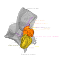
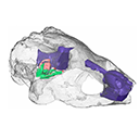
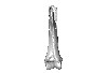
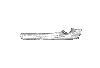
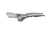
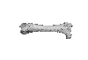
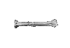
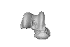
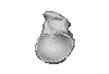
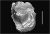
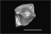
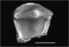
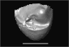
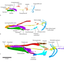
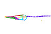
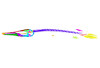
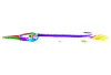
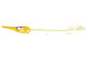
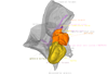
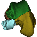
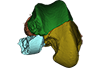
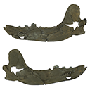
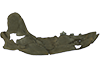
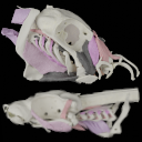
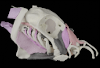
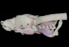
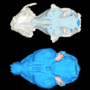
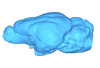
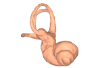
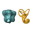
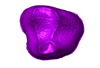
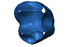
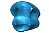
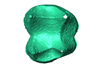
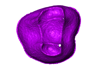
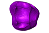
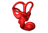
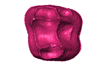
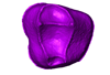
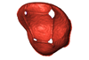
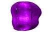
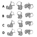
This project presents the osteological connexions of the petrosal bone of the extant Hippopotamidae Hippopotamus amphibius and Choeropsis liberiensis by a virtual osteological dissection of the ear region. The petrosal, the bulla, the sinuses and the major morphological features surrounding the petrosal bone are labelled, both in situ and in an exploded model presenting disassembly views. The directional underwater hearing mode of Hippopotamidae is discussed based on the new observations.
Choeropsis liberiensis UPPal-M09-5-005a View specimen
|
M3#1Labelled compact model of the right ear region of Choeropsis liberiensis (UPPal-M09-5-005a) Type: "3D_surfaces"doi: 10.18563/m3.sf1 state:published |
Download 3D surface file |

|
M3#2Labelled exploded model of the right ear region of Choeropsis liberiensis (UPPal-M09-5-005a) Type: "3D_surfaces"doi: 10.18563/m3.sf2 state:published |
Download 3D surface file |
Hippopotamus amphibius UM N179 View specimen

|
M3#3Labelled compact model of the right ear region of Hippopotamus amphibius (UM N 179) Type: "3D_surfaces"doi: 10.18563/m3.sf3 state:published |
Download 3D surface file |

|
M3#4Labelled exploded model of the right ear region of Hippopotamus amphibius (UM N 179) Type: "3D_surfaces"doi: 10.18563/m3.sf4 state:published |
Download 3D surface file |
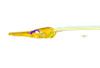
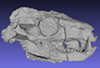
The present 3D Dataset contains the 3D model analyzed in The largest freshwater odontocete: a South Asian river dolphin relative from the Proto-Amazonia.
Pebanista yacuruna MUSM 4017 View specimen
|
M3#1394Holotype skull of Pebanista yacuruna MUSM 4017 Type: "3D_surfaces"doi: 10.18563/m3.sf.1394 state:published |
Download 3D surface file |
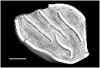
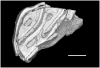
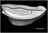
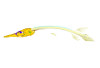
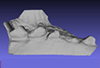
The present contribution contains the 3D virtual restoration of a Pliocene Lutrine right femur of Tobène, Senegal, described and figured in Lihoreau et al. (2021) : "A fossil terrestrial fauna from Tobène (Senegal) provides a unique early Pliocene window in Western Africa ". https://doi.org/10.1016/j.gr.2021.06.013
Indet indet SN-Tob-12-02 View specimen
|
M3#441Virtual restoration of SN-Tob-12-02 Type: "3D_surfaces"doi: 10.18563/m3.sf.441 state:published |
Download 3D surface file |
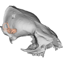
The present 3D Dataset contains 3D models of the cranium surface and of the bony labyrinth endocast of the stem bat Vielasia sigei. They are used by (Hand et al., 2023) to explore the phylogenetic position of this species, to infer its laryngeal echolocating capabilities, and to eventually discuss chiropteran evolution before the crown clade diversification.
Vielasia sigei UM VIE-250 View specimen
|
M3#1269External surface of the cranium Type: "3D_surfaces"doi: 10.18563/m3.sf.1269 state:published |
Download 3D surface file |
|
M3#1270Virtual endocast of the right bony labyrinth Type: "3D_surfaces"doi: 10.18563/m3.sf.1270 state:published |
Download 3D surface file |
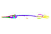
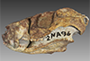
The present 3D Dataset contains the 3D model analyzed in Solé F., Lesport J.-F., Heitz A., and Mennecart B. minor revision. A new gigantic carnivore (Carnivora, Amphicyonidae) from the late middle Miocene of France. PeerJ.
Tartarocyon cazanavei MHNBx 2020.20.1 View specimen
|
M3#903Surface scan (ply) and texture (png) of the holotype of Tartarocyon cazanavei (MHNBx 2020.20.1) Type: "3D_surfaces"doi: 10.18563/m3.sf.903 state:published |
Download 3D surface file |
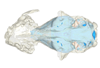
This contribution contains 3D models of the cranial skeleton and muscles in an elephantfish (Callorhinchus milii) and a catshark (Scyliorhinus canicula), based on synchrotron tomographic scans. These datasets were analyzed and described in Dearden et al. (2021) “The morphology and evolution of chondrichthyan cranial muscles: a digital dissection of the elephantfish Callorhinchus milii and the catshark Scyliorhinus canicula.” Journal of Anatomy.
Callorhinchus milii 001 View specimen
|
M3#7083D models of the cranial skeleton and muscles of Callorhinchus milii, created using Mimics. Type: "3D_surfaces"doi: 10.18563/m3.sf.708 state:published |
Download 3D surface file |
Scyliorhinus canicula 002 View specimen
|
M3#7093D models of the cranial skeleton and muscles of Scyliorhinus canicula, created using Mimics. Type: "3D_surfaces"doi: 10.18563/m3.sf.709 state:published |
Download 3D surface file |
The present 3D Dataset contains the 3D model of the brain endocast of Neoepiblema acreensis analyzed in “Small within the largest: Brain size and anatomy of the extinct Neoepiblema acreensis, a giant rodent from the Neotropics”. The 3D model was generated using CT-Scanning and techniques of virtual reconstruction.
Neoepiblema acreensis UFAC 4515 View specimen
|
M3#502Brain endocast of Neoepiblema acreensis Type: "3D_surfaces"doi: 10.18563/m3.sf.502 state:published |
Download 3D surface file |

This contribution contains the 3D models described and figured in the following publication: Molnar, JL, Pierce, SE, Bhullar, B-A, Turner, AH, Hutchinson, JR (accepted). Morphological and functional changes in the crocodylomorph vertebral column with increasing aquatic adaptation. Royal Society Open Science.
Protosuchus richardsoni AMNH-VP 3024 View specimen

|
M3#448th and 9th dorsal vertebrae, 1st and 2nd lumbar vertebrae, and 5th lumbar and sacral vertebrae. Type: "3D_surfaces"doi: 10.18563/m3.sf44 state:published |
Download 3D surface file |
Terrestrisuchus gracilis NHM-PV R 7562 View specimen

|
M3#451st and 2nd lumbar vertebrae, and 5th lumbar and sacral vertebrae Type: "3D_surfaces"doi: 10.18563/m3.sf45 state:published |
Download 3D surface file |
Pelagosaurus typus NHM-PV OR 32598 View specimen

|
M3#467th and 8th dorsal vertebrae, 11th and 12th dorsal vertebrae, 15th dorsal vertebra and sacral vertebra. Type: "3D_surfaces"doi: 10.18563/m3.sf46 state:published |
Download 3D surface file |
Metriorhynchus superciliosus NHM-PV R 2054 View specimen

|
M3#476th and 7th dorsal vertebrae, 10th and 11th dorsal vertebrae, 17th dorsal vertebra and sacral vertebra Type: "3D_surfaces"doi: 10.18563/m3.sf47 state:published |
Download 3D surface file |
Crocodylus niloticus FNC0 View specimen

|
M3#487th and 8th dorsal vertebrae, 1st and 2nd lumbar vertebrae, 5th lumbar vertebra and sacral vertebra. Type: "3D_surfaces"doi: 10.18563/m3.sf48 state:published |
Download 3D surface file |
This contribution contains the 3D models described and figured in the following publication: Bonis, L. de, Grohé, C., Surault, J., Gardin, A. 2022. Description of the first cranium and endocranial structures of Stenoplesictis minor (Mammalia, Carnivora), an early aeluroid from the Oligocene of the Quercy Phosphorites (southwestern France). Historical Biology. https://doi.org/10.1080/08912963.2022.2045980
Stenoplesictis minor UM-ACQ 6705 View specimen
|
M3#961Endocranium Type: "3D_surfaces"doi: 10.18563/m3.sf.961 state:published |
Download 3D surface file |
|
M3#962Right bony labyrinth Type: "3D_surfaces"doi: 10.18563/m3.sf.962 state:published |
Download 3D surface file |

|
M3#963Left bony labyrinth Type: "3D_surfaces"doi: 10.18563/m3.sf.963 state:published |
Download 3D surface file |
|
M3#964Cranium in transparency with endocranial structures Type: "3D_surfaces"doi: 10.18563/m3.sf.964 state:published |
Download 3D surface file |
The present 3D Dataset contains the 3D models analyzed in Hendrickx, C., Gaetano, L. C., Choiniere, J., Mocke, H. and Abdala, F. in press. A new traversodontid cynodont with a peculiar postcanine dentition from the Middle/Late Triassic of Namibia and dental evolution in basal gomphodonts. Journal of Systematic Palaeontology.
Etjoia dentitransitus GSN F1591 View specimen
|
M3#557Surface model derived from µCT data of the holotype of Etjoia dentitransitus Type: "3D_surfaces"doi: 10.18563/m3.sf.557 state:published |
Download 3D surface file |
|
M3#558Photogrammetric 3D surface model of the postcanines of the Holotype of Etjoia dentitransitus Type: "3D_surfaces"doi: 10.18563/m3.sf.558 state:published |
Download 3D surface file |
|
M3#559Photogrammetric 3D surface model of the Holotype of Etjoia dentitransitus Type: "3D_surfaces"doi: 10.18563/m3.sf.559 state:published |
Download 3D surface file |
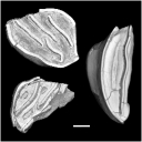
This contribution contains the 3D models of the ossicles of a protocetid archaeocete from the locality of Kpogamé, Togo, described and figured in the publication of Mourlam and Orliac (2019).
indet. indet. UM KPG-M 73 View specimen

|
M3#407stapes Type: "3D_surfaces"doi: 10.18563/m3.sf.407 state:published |
Download 3D surface file |

|
M3#408Incus Type: "3D_surfaces"doi: 10.18563/m3.sf.408 state:published |
Download 3D surface file |

|
M3#409Malleus Type: "3D_surfaces"doi: 10.18563/m3.sf.409 state:published |
Download 3D surface file |
The present 3D Dataset contains the 3D models of the enamel-dentine junctions of upper third molars and of the bony labyrinths of the extant cercopithecoid specimens analyzed in the following publication: Beaudet, A., Dumoncel, J., Thackeray, J.F., Bruxelles, L., Duployer, B., Tenailleau, C., Bam, L., Hoffman, J., de Beer, F., Braga, J.: Upper third molar internal structural organization and semicircular canal morphology in Plio-Pleistocene South African cercopithecoids. Journal of Human Evolution 95, 104-120. https://doi.org/10.1016/j.jhevol.2016.04.004
Cercocebus atys 81.007-M-0041 View specimen
|
M3#4453D model of the enamel-dentine junction of the right upper third molar. Type: "3D_surfaces"doi: 10.18563/m3.sf.445 state:published |
Download 3D surface file |
Cercocebus torquatus 73.018-M-0359 View specimen
|
M3#4463D model of the enamel-dentine junction of the right upper third molar. Type: "3D_surfaces"doi: 10.18563/m3.sf.446 state:published |
Download 3D surface file |

|
M3#4963D model of the left bony labyrinth. Type: "3D_surfaces"doi: 10.18563/m3.sf.496 state:published |
Download 3D surface file |
Mandrillus leucophaeus 73.029-M-0106 View specimen
|
M3#4473D model of the enamel-dentine junction of the right upper third molar. Type: "3D_surfaces"doi: 10.18563/m3.sf.447 state:published |
Download 3D surface file |

|
M3#4703D model of the right bony labyrinth. Type: "3D_surfaces"doi: 10.18563/m3.sf.470 state:published |
Download 3D surface file |
Lophocebus albigena 73.029-M-0109 View specimen
|
M3#4483D model of the enamel-dentine junction of the right upper third molar. Type: "3D_surfaces"doi: 10.18563/m3.sf.448 state:published |
Download 3D surface file |

|
M3#4713D model of the right bony labyrinth. Type: "3D_surfaces"doi: 10.18563/m3.sf.471 state:published |
Download 3D surface file |
Piliocolobus foai 91.060-M-0071 View specimen
|
M3#4493D model of the enamel-dentine junction of the right upper third molar. Type: "3D_surfaces"doi: 10.18563/m3.sf.449 state:published |
Download 3D surface file |

|
M3#4723D model of the right bony labyrinth. Type: "3D_surfaces"doi: 10.18563/m3.sf.472 state:published |
Download 3D surface file |
Colobus guereza 1215 View specimen
|
M3#4503D model of the enamel-dentine junction of the right upper third molar. Type: "3D_surfaces"doi: 10.18563/m3.sf.450 state:published |
Download 3D surface file |

|
M3#4733D model of the right bony labyrinth. Type: "3D_surfaces"doi: 10.18563/m3.sf.473 state:published |
Download 3D surface file |
Colobus guereza 2800 View specimen
|
M3#4513D model of the enamel-dentine junction of the right upper third molar. Type: "3D_surfaces"doi: 10.18563/m3.sf.451 state:published |
Download 3D surface file |

|
M3#4743D model of the right bony labyrinth. Type: "3D_surfaces"doi: 10.18563/m3.sf.474 state:published |
Download 3D surface file |
Papio cynocephalus kindae 3503 View specimen
|
M3#4523D model of the enamel-dentine junction of the right upper third molar. Type: "3D_surfaces"doi: 10.18563/m3.sf.452 state:published |
Download 3D surface file |

|
M3#4753D model of the right bony labyrinth. Type: "3D_surfaces"doi: 10.18563/m3.sf.475 state:published |
Download 3D surface file |
Erythrocebus patas 8452 View specimen
|
M3#4533D model of the enamel-dentine junction of the right upper third molar. Type: "3D_surfaces"doi: 10.18563/m3.sf.453 state:published |
Download 3D surface file |

|
M3#4763D model of the right bony labyrinth. Type: "3D_surfaces"doi: 10.18563/m3.sf.476 state:published |
Download 3D surface file |
Papio cynocephalus kindae 17979 View specimen
|
M3#4543D model of the enamel-dentine junction of the right upper third molar. Type: "3D_surfaces"doi: 10.18563/m3.sf.454 state:published |
Download 3D surface file |

|
M3#4773D model of the right bony labyrinth. Type: "3D_surfaces"doi: 10.18563/m3.sf.477 state:published |
Download 3D surface file |
Colobus angolensis 25456 View specimen
|
M3#4553D model of the enamel-dentine junction of the right upper third molar. Type: "3D_surfaces"doi: 10.18563/m3.sf.455 state:published |
Download 3D surface file |

|
M3#4783D model of the right bony labyrinth. Type: "3D_surfaces"doi: 10.18563/m3.sf.478 state:published |
Download 3D surface file |
Chlorocebus pygerythrus 37477 View specimen
|
M3#4563D model of the enamel-dentine junction of the right upper third molar. Type: "3D_surfaces"doi: 10.18563/m3.sf.456 state:published |
Download 3D surface file |
|
M3#4813D model of the right bony labyrinth. Type: "3D_surfaces"doi: 10.18563/m3.sf.481 state:published |
Download 3D surface file |
Chlorocebus pygerythrus 37478 View specimen
|
M3#4573D model of the enamel-dentine junction of the right upper third molar. Type: "3D_surfaces"doi: 10.18563/m3.sf.457 state:published |
Download 3D surface file |

|
M3#4823D model of the right bony labyrinth. Type: "3D_surfaces"doi: 10.18563/m3.sf.482 state:published |
Download 3D surface file |
Lophocebus albigena 37572 View specimen
|
M3#4583D model of the enamel-dentine junction of the right upper third molar. Type: "3D_surfaces"doi: 10.18563/m3.sf.458 state:published |
Download 3D surface file |

|
M3#4833D model of the right bony labyrinth. Type: "3D_surfaces"doi: 10.18563/m3.sf.483 state:published |
Download 3D surface file |
Lophocebus albigena 37579 View specimen
|
M3#4593D model of the enamel-dentine junction of the right upper third molar. Type: "3D_surfaces"doi: 10.18563/m3.sf.459 state:published |
Download 3D surface file |
Erythrocebus patas OST.2002-26 View specimen
|
M3#4603D model of the enamel-dentine junction of the right upper third molar. Type: "3D_surfaces"doi: 10.18563/m3.sf.460 state:published |
Download 3D surface file |
|
M3#4843D model of the right bony labyrinth. Type: "3D_surfaces"doi: 10.18563/m3.sf.484 state:published |
Download 3D surface file |
Mandrillus sphinx OST.AC.488 View specimen
|
M3#4613D model of the enamel-dentine junction of the right upper third molar. Type: "3D_surfaces"doi: 10.18563/m3.sf.461 state:published |
Download 3D surface file |

|
M3#4853D model of the left bony labyrinth. Type: "3D_surfaces"doi: 10.18563/m3.sf.485 state:published |
Download 3D surface file |
Macaca mulatta OST.AC.492 View specimen
|
M3#4623D model of the enamel-dentine junction of the right upper third molar. Type: "3D_surfaces"doi: 10.18563/m3.sf.462 state:published |
Download 3D surface file |

|
M3#4863D model of the right bony labyrinth. Type: "3D_surfaces"doi: 10.18563/m3.sf.486 state:published |
Download 3D surface file |
Chlorocebus aethiops OST.AC.523 View specimen
|
M3#4633D model of the enamel-dentine junction of the right upper third molar. Type: "3D_surfaces"doi: 10.18563/m3.sf.463 state:published |
Download 3D surface file |

|
M3#4913D model of the right bony labyrinth. Type: "3D_surfaces"doi: 10.18563/m3.sf.491 state:published |
Download 3D surface file |
Cercopithecus cephus OST.AC.533 View specimen
|
M3#4643D model of the enamel-dentine junction of the right upper third molar. Type: "3D_surfaces"doi: 10.18563/m3.sf.464 state:published |
Download 3D surface file |

|
M3#4933D model of the right bony labyrinth. Type: "3D_surfaces"doi: 10.18563/m3.sf.493 state:published |
Download 3D surface file |
Chlorocebus aethiops OST.AC.540 View specimen
|
M3#4653D model of the enamel-dentine junction of the right upper third molar. Type: "3D_surfaces"doi: 10.18563/m3.sf.465 state:published |
Download 3D surface file |

|
M3#4943D model of the right bony labyrinth. Type: "3D_surfaces"doi: 10.18563/m3.sf.494 state:published |
Download 3D surface file |
Mandrillus sphinx OST.AC.543 View specimen
|
M3#4663D model of the enamel-dentine junction of the right upper third molar. Type: "3D_surfaces"doi: 10.18563/m3.sf.466 state:published |
Download 3D surface file |
|
M3#4953D model of the right bony labyrinth. Type: "3D_surfaces"doi: 10.18563/m3.sf.495 state:published |
Download 3D surface file |
Cercocebus torquatus 73.018-M-389 View specimen

|
M3#4683D model of the right bony labyrinth. Type: "3D_surfaces"doi: 10.18563/m3.sf.468 state:published |
Download 3D surface file |
Mandrillus leucophaeus 73.029-M-0105 View specimen
|
M3#4693D model of the right bony labyrinth. Type: "3D_surfaces"doi: 10.18563/m3.sf.469 state:published |
Download 3D surface file |
Mandrillus leucophaeus 28425 View specimen

|
M3#4793D model of the right bony labyrinth. Type: "3D_surfaces"doi: 10.18563/m3.sf.479 state:published |
Download 3D surface file |
Cercocebus atys 28998 View specimen

|
M3#4803D model of the right bony labyrinth. Type: "3D_surfaces"doi: 10.18563/m3.sf.480 state:published |
Download 3D surface file |
Macaca sylvanus OST.AC.493 View specimen

|
M3#4873D model of the right bony labyrinth. Type: "3D_surfaces"doi: 10.18563/m3.sf.487 state:published |
Download 3D surface file |
Chlorocebus aethiops OST.AC.508 View specimen

|
M3#4883D model of the left bony labyrinth. Type: "3D_surfaces"doi: 10.18563/m3.sf.488 state:published |
Download 3D surface file |
Cercopithecus cephus OST.AC.515 View specimen

|
M3#4893D model of the right bony labyrinth. Type: "3D_surfaces"doi: 10.18563/m3.sf.489 state:published |
Download 3D surface file |
Colobus guereza OST.AC.519 View specimen

|
M3#4903D model of the right bony labyrinth. Type: "3D_surfaces"doi: 10.18563/m3.sf.490 state:published |
Download 3D surface file |
Macaca sp. OST.AC.532 View specimen

|
M3#4923D model of the left bony labyrinth. Type: "3D_surfaces"doi: 10.18563/m3.sf.492 state:published |
Download 3D surface file |
The present 3D Dataset contains the 3D models analyzed in Velazco P. M., Grohé C. 2017. Comparative anatomy of the bony labyrinth of the bats Platalina genovensium (Phyllostomidae, Lonchophyllinae) and Tomopeas ravus (Molossidae, Tomopeatinae). Biotempo 14(2).
Platalina genovensium 278520 View specimen

|
M3#276Right bony labyrinth surface positioned (.PLY) Labels associated (.FLG) Type: "3D_surfaces"doi: 10.18563/m3.sf.276 state:published |
Download 3D surface file |
Tomopeas ravus 278525 View specimen

|
M3#277Right bony labyrinth surface (.PLY) Labels associated (.FLG) Type: "3D_surfaces"doi: 10.18563/m3.sf.277 state:published |
Download 3D surface file |
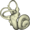
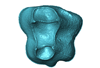
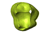
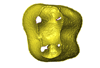
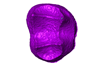
The present 3D Dataset contains the 3D models analyzed in the following publication: Size variation under domestication: Conservatism in the inner ear shape of wolves, dogs and dingoes. Scientific Reports 7, Article number: 13330, https://doi.org/10.1038/s41598-017-13523-9.
Canis lupus familiaris NMBE 16 View specimen

|
M3#2293D virtual endocast of the left inner ear Type: "3D_surfaces"doi: 10.18563/m3.sf.229 state:published |
Download 3D surface file |
Canis lupus familiaris NMBE-LAT-1136 View specimen

|
M3#2423D virtual endocast of the left inner ear Type: "3D_surfaces"doi: 10.18563/m3.sf.242 state:published |
Download 3D surface file |
Canis lupus familiaris NMBE-LAT-1119 View specimen

|
M3#2433D virtual endocast of the left inner ear Type: "3D_surfaces"doi: 10.18563/m3.sf.243 state:published |
Download 3D surface file |
Canis lupus familiaris NMBE-BUR-1057 View specimen

|
M3#2443D virtual endocast of the left inner ear Type: "3D_surfaces"doi: 10.18563/m3.sf.244 state:published |
Download 3D surface file |
Canis lupus familiaris NMBE-LUS-1102 View specimen

|
M3#2453D virtual endocast of the left inner ear Type: "3D_surfaces"doi: 10.18563/m3.sf.245 state:published |
Download 3D surface file |
Canis lupus familiaris NMBE-LUS-1095 View specimen

|
M3#2463D virtual endocast of the left inner ear Type: "3D_surfaces"doi: 10.18563/m3.sf.246 state:published |
Download 3D surface file |
Canis lupus familiaris NMBE-DUR-1124 View specimen

|
M3#2473D virtual endocast of the left inner ear Type: "3D_surfaces"doi: 10.18563/m3.sf.247 state:published |
Download 3D surface file |
Canis lupus chanco ZMUZH 17603 View specimen

|
M3#2483D virtual endocast of the left inner ear Type: "3D_surfaces"doi: 10.18563/m3.sf.248 state:published |
Download 3D surface file |
Canis lupus chanco ZMUZH 20201 View specimen

|
M3#2493D virtual endocast of the left inner ear Type: "3D_surfaces"doi: 10.18563/m3.sf.249 state:published |
Download 3D surface file |
Canis lupus chanco ZMUZH 17602 View specimen

|
M3#2503D virtual endocast of the left inner ear Type: "3D_surfaces"doi: 10.18563/m3.sf.250 state:published |
Download 3D surface file |
Canis lupus ZMUZH 13854 View specimen

|
M3#2403D virtual endocast of the left inner ear Type: "3D_surfaces"doi: 10.18563/m3.sf.240 state:published |
Download 3D surface file |
Canis lupus chanco ZMUZH 20202 View specimen

|
M3#2393D virtual endocast of the left inner ear Type: "3D_surfaces"doi: 10.18563/m3.sf.239 state:published |
Download 3D surface file |
Canis lupus chanco ZMUZH 17612 View specimen

|
M3#2303D virtual endocast of the left inner ear Type: "3D_surfaces"doi: 10.18563/m3.sf.230 state:published |
Download 3D surface file |
Canis lupus chanco ZMUZH 18082 View specimen

|
M3#2313D virtual endocast of the left inner ear Type: "3D_surfaces"doi: 10.18563/m3.sf.231 state:published |
Download 3D surface file |
Canis lupus ZMUZH 17118 View specimen

|
M3#2323D virtual endocast of the left inner ear Type: "3D_surfaces"doi: 10.18563/m3.sf.232 state:published |
Download 3D surface file |
Canis lupus ZMUZH 15858 View specimen

|
M3#2333D virtual endocast of the left inner ear Type: "3D_surfaces"doi: 10.18563/m3.sf.233 state:published |
Download 3D surface file |
Canis lupus familiaris ZMUZH 17712 View specimen

|
M3#2343D virtual endocast of the left inner ear Type: "3D_surfaces"doi: 10.18563/m3.sf.234 state:published |
Download 3D surface file |
Canis lupus familiaris ZMUZH 17713 View specimen

|
M3#2353D virtual endocast of the left inner ear Type: "3D_surfaces"doi: 10.18563/m3.sf.235 state:published |
Download 3D surface file |
Canis lupus familiaris ZMUZH 10166 View specimen

|
M3#2363D virtual endocast of the left inner ear Type: "3D_surfaces"doi: 10.18563/m3.sf.236 state:published |
Download 3D surface file |
Canis lupus familiaris ZMUZH 10175 View specimen

|
M3#2373D virtual endocast of the left inner ear Type: "3D_surfaces"doi: 10.18563/m3.sf.237 state:published |
Download 3D surface file |
Canis lupus familiaris ZMUZH 14842 View specimen

|
M3#2383D virtual endocast of the left inner ear Type: "3D_surfaces"doi: 10.18563/m3.sf.238 state:published |
Download 3D surface file |
Canis lupus familiaris ZMUZH 10342 View specimen

|
M3#2513D virtual endocast of the left inner ear Type: "3D_surfaces"doi: 10.18563/m3.sf.251 state:published |
Download 3D surface file |
Canis lupus familiaris ZMUZH 10343 View specimen

|
M3#2523D virtual endocast of the left inner ear Type: "3D_surfaces"doi: 10.18563/m3.sf.252 state:published |
Download 3D surface file |
Canis lupus familiaris ZMUZH 13766 View specimen

|
M3#2533D virtual endocast of the left inner ear Type: "3D_surfaces"doi: 10.18563/m3.sf.253 state:published |
Download 3D surface file |
Canis lupus familiaris ZMUZH 17717 View specimen

|
M3#2653D virtual endocast of the left inner ear Type: "3D_surfaces"doi: 10.18563/m3.sf.265 state:published |
Download 3D surface file |
Canis lupus familiaris ZMUZH 17711 View specimen

|
M3#2663D virtual endocast of the left inner ear Type: "3D_surfaces"doi: 10.18563/m3.sf.266 state:published |
Download 3D surface file |
Canis lupus familiaris ZMUZH 17714 View specimen

|
M3#2673D virtual endocast of the left inner ear Type: "3D_surfaces"doi: 10.18563/m3.sf.267 state:published |
Download 3D surface file |
Canis lupus familiaris ZMUZH 17715 View specimen

|
M3#2683D virtual endocast of the left inner ear Type: "3D_surfaces"doi: 10.18563/m3.sf.268 state:published |
Download 3D surface file |
Canis lupus familiaris PIMUZ A/V 2835 View specimen

|
M3#2693D virtual endocast of the left inner ear Type: "3D_surfaces"doi: 10.18563/m3.sf.269 state:published |
Download 3D surface file |
Canis lupus familiaris PIMUZ A/V 2834 View specimen

|
M3#2703D virtual endocast of the left inner ear Type: "3D_surfaces"doi: 10.18563/m3.sf.270 state:published |
Download 3D surface file |
Canis lupus familiaris PIMUZ A/V 2837 View specimen

|
M3#2713D virtual endocast of the left inner ear Type: "3D_surfaces"doi: 10.18563/m3.sf.271 state:published |
Download 3D surface file |
Canis lupus familiaris PIMUZ A/V 2831 View specimen

|
M3#2723D virtual endocast of the left inner ear Type: "3D_surfaces"doi: 10.18563/m3.sf.272 state:published |
Download 3D surface file |
Canis lupus familiaris PIMUZ A/V 2845 View specimen

|
M3#2733D virtual endocast of the left inner ear Type: "3D_surfaces"doi: 10.18563/m3.sf.273 state:published |
Download 3D surface file |
Canis lupus familiaris PIMUZ A/V 3001 View specimen

|
M3#2643D virtual endocast of the left inner ear Type: "3D_surfaces"doi: 10.18563/m3.sf.264 state:published |
Download 3D surface file |
Canis lupus familiaris PIMUZ A/V 2832 View specimen

|
M3#2633D virtual endocast of the left inner ear Type: "3D_surfaces"doi: 10.18563/m3.sf.263 state:published |
Download 3D surface file |
Canis lupus familiaris PIMUZ A/V 3000 View specimen

|
M3#2543D virtual endocast of the left inner ear Type: "3D_surfaces"doi: 10.18563/m3.sf.254 state:published |
Download 3D surface file |
Canis lupus familiaris PIMUZ A/V 2847 View specimen

|
M3#2553D virtual endocast of the left inner ear Type: "3D_surfaces"doi: 10.18563/m3.sf.255 state:published |
Download 3D surface file |
Canis lupus familiaris PIMUZ A/V 2846 View specimen

|
M3#2563D virtual endocast of the left inner ear Type: "3D_surfaces"doi: 10.18563/m3.sf.256 state:published |
Download 3D surface file |
Canis lupus familiaris PIMUZ A/V 2836 View specimen

|
M3#2573D virtual endocast of the left inner ear Type: "3D_surfaces"doi: 10.18563/m3.sf.257 state:published |
Download 3D surface file |
Canis lupus familiaris NMB 12080 View specimen

|
M3#2583D virtual endocast of the left inner ear Type: "3D_surfaces"doi: 10.18563/m3.sf.258 state:published |
Download 3D surface file |
Canis lupus familiaris NMB 12081 View specimen

|
M3#2593D virtual endocast of the left inner ear Type: "3D_surfaces"doi: 10.18563/m3.sf.259 state:published |
Download 3D surface file |
Canis lupus familiaris NMB 12079 View specimen

|
M3#2603D virtual endocast of the left inner ear Type: "3D_surfaces"doi: 10.18563/m3.sf.260 state:published |
Download 3D surface file |
Canis lupus familiaris NMB 12078 View specimen

|
M3#2613D virtual endocast of the left inner ear Type: "3D_surfaces"doi: 10.18563/m3.sf.261 state:published |
Download 3D surface file |
Canis lupus familiaris NMBE 1051209 View specimen

|
M3#2623D virtual endocast of the left inner ear Type: "3D_surfaces"doi: 10.18563/m3.sf.262 state:published |
Download 3D surface file |
Canis lupus familiaris NMBE 1051226 View specimen

|
M3#2283D virtual endocast of the left inner ear Type: "3D_surfaces"doi: 10.18563/m3.sf.228 state:published |
Download 3D surface file |
Canis lupus familiaris NMBE 1051381 View specimen

|
M3#2213D virtual endocast of the left inner ear Type: "3D_surfaces"doi: 10.18563/m3.sf.221 state:published |
Download 3D surface file |
Canis lupus familiaris NMBE 1051418 View specimen

|
M3#1843D virtual endocast of the left inner ear Type: "3D_surfaces"doi: 10.18563/m3.sf.184 state:published |
Download 3D surface file |
Canis lupus familiaris ZMUZH A.II. View specimen

|
M3#1973D virtual endocast of the left inner ear Type: "3D_surfaces"doi: 10.18563/m3.sf.197 state:published |
Download 3D surface file |
Canis lupus familiaris ZMUZH A.VII. View specimen

|
M3#1983D virtual endocast of the left inner ear Type: "3D_surfaces"doi: 10.18563/m3.sf.198 state:published |
Download 3D surface file |
Canis lupus familiaris ZMUZH We.6. View specimen

|
M3#1993D virtual endocast of the left inner ear Type: "3D_surfaces"doi: 10.18563/m3.sf.199 state:published |
Download 3D surface file |
Canis lupus familiaris ZMUZH Ez.2. View specimen

|
M3#2003D virtual endocast of the left inner ear Type: "3D_surfaces"doi: 10.18563/m3.sf.200 state:published |
Download 3D surface file |
Canis lupus familiaris ZMUZH Ez.E. View specimen

|
M3#2013D virtual endocast of the left inner ear Type: "3D_surfaces"doi: 10.18563/m3.sf.201 state:published |
Download 3D surface file |
Canis lupus familiaris ZMUZH A.6. View specimen

|
M3#2023D virtual endocast of the left inner ear Type: "3D_surfaces"doi: 10.18563/m3.sf.202 state:published |
Download 3D surface file |
Canis lupus familiaris ZMUZH Wyn.9. View specimen

|
M3#2033D virtual endocast of the left inner ear Type: "3D_surfaces"doi: 10.18563/m3.sf.203 state:published |
Download 3D surface file |
Canis lupus familiaris ZMUZH F.48. View specimen

|
M3#2043D virtual endocast of the left inner ear Type: "3D_surfaces"doi: 10.18563/m3.sf.204 state:published |
Download 3D surface file |
Canis lupus familiaris ZMUZH Terp.1. View specimen

|
M3#2053D virtual endocast of the left inner ear Type: "3D_surfaces"doi: 10.18563/m3.sf.205 state:published |
Download 3D surface file |
Canis lupus familiaris ZMUZH A.VIII. View specimen

|
M3#1963D virtual endocast of the left inner ear Type: "3D_surfaces"doi: 10.18563/m3.sf.196 state:published |
Download 3D surface file |
Canis lupus familiaris ZMUZH A.VI. View specimen

|
M3#1953D virtual endocast of the left inner ear Type: "3D_surfaces"doi: 10.18563/m3.sf.195 state:published |
Download 3D surface file |
Canis lupus familiaris ZMUZH A.IV. View specimen

|
M3#1853D virtual endocast of the left inner ear Type: "3D_surfaces"doi: 10.18563/m3.sf.185 state:published |
Download 3D surface file |
Canis lupus familiaris NMBE A.403. View specimen

|
M3#1873D virtual endocast of the left inner ear Type: "3D_surfaces"doi: 10.18563/m3.sf.187 state:published |
Download 3D surface file |
Canis lupus familiaris NMBE A.5.a. View specimen

|
M3#1883D virtual endocast of the left inner ear Type: "3D_surfaces"doi: 10.18563/m3.sf.188 state:published |
Download 3D surface file |
Canis lupus NMB 8381 View specimen

|
M3#1893D virtual endocast of the left inner ear Type: "3D_surfaces"doi: 10.18563/m3.sf.189 state:published |
Download 3D surface file |
Canis lupus lycaon NMB C.1362 View specimen

|
M3#1903D virtual endocast of the left inner ear Type: "3D_surfaces"doi: 10.18563/m3.sf.190 state:published |
Download 3D surface file |
Canis lupus NMB Z309 View specimen

|
M3#1913D virtual endocast of the left inner ear Type: "3D_surfaces"doi: 10.18563/m3.sf.191 state:published |
Download 3D surface file |
Canis lupus NMB 2761 View specimen

|
M3#1923D virtual endocast of the left inner ear Type: "3D_surfaces"doi: 10.18563/m3.sf.192 state:published |
Download 3D surface file |
Canis lupus occidentalis NMB No Nb View specimen

|
M3#1933D virtual endocast of the left inner ear Type: "3D_surfaces"doi: 10.18563/m3.sf.193 state:published |
Download 3D surface file |
Canis lupus NMB 5258 View specimen

|
M3#1943D virtual endocast of the left inner ear Type: "3D_surfaces"doi: 10.18563/m3.sf.194 state:published |
Download 3D surface file |
Canis lupus NMB SCM320 View specimen

|
M3#2063D virtual endocast of the left inner ear Type: "3D_surfaces"doi: 10.18563/m3.sf.206 state:published |
Download 3D surface file |
Canis lupus arabs NMB 11019 View specimen

|
M3#2073D virtual endocast of the left inner ear Type: "3D_surfaces"doi: 10.18563/m3.sf.207 state:published |
Download 3D surface file |
Canis lupus UMZC K.3141 View specimen

|
M3#2083D virtual endocast of the left inner ear Type: "3D_surfaces"doi: 10.18563/m3.sf.208 state:published |
Download 3D surface file |
Canis lupus UMZC K.3150.1 View specimen

|
M3#2193D virtual endocast of the left inner ear Type: "3D_surfaces"doi: 10.18563/m3.sf.219 state:published |
Download 3D surface file |
Canis lupus UMZC K.3152 View specimen

|
M3#2203D virtual endocast of the left inner ear Type: "3D_surfaces"doi: 10.18563/m3.sf.220 state:published |
Download 3D surface file |
Canis lupus UMZC K.3149 View specimen

|
M3#2223D virtual endocast of the left inner ear Type: "3D_surfaces"doi: 10.18563/m3.sf.222 state:published |
Download 3D surface file |
Canis lupus familiaris UMZC K.3016 View specimen

|
M3#2233D virtual endocast of the left inner ear Type: "3D_surfaces"doi: 10.18563/m3.sf.223 state:published |
Download 3D surface file |
Canis lupus occidentalis ZMUZH 17210 View specimen

|
M3#2243D virtual endocast of the left inner ear Type: "3D_surfaces"doi: 10.18563/m3.sf.224 state:published |
Download 3D surface file |
Canis lupus familiaris SZ 7961 View specimen

|
M3#2253D virtual endocast of the left inner ear Type: "3D_surfaces"doi: 10.18563/m3.sf.225 state:published |
Download 3D surface file |
Canis lupus familiaris SZ 7959 View specimen

|
M3#2263D virtual endocast of the left inner ear Type: "3D_surfaces"doi: 10.18563/m3.sf.226 state:published |
Download 3D surface file |
Canis lupus familiaris SZ 7958 View specimen

|
M3#2173D virtual endocast of the left inner ear Type: "3D_surfaces"doi: 10.18563/m3.sf.217 state:published |
Download 3D surface file |
Canis lupus familiaris SZ 7930 View specimen

|
M3#2163D virtual endocast of the left inner ear Type: "3D_surfaces"doi: 10.18563/m3.sf.216 state:published |
Download 3D surface file |
Canis lupus familiaris SZ 7926 View specimen

|
M3#2183D virtual endocast of the left inner ear Type: "3D_surfaces"doi: 10.18563/m3.sf.218 state:published |
Download 3D surface file |
Canis lupus familiaris SZ 7929 View specimen

|
M3#2093D virtual endocast of the left inner ear Type: "3D_surfaces"doi: 10.18563/m3.sf.209 state:published |
Download 3D surface file |
Canis lupus dingo M6297 View specimen

|
M3#1863D virtual endocast of the left inner ear Type: "3D_surfaces"doi: 10.18563/m3.sf.186 state:published |
Download 3D surface file |
Canis lupus dingo M24153 View specimen

|
M3#2103D virtual endocast of the left inner ear Type: "3D_surfaces"doi: 10.18563/m3.sf.210 state:published |
Download 3D surface file |
Canis lupus dingo M33608 View specimen

|
M3#2113D virtual endocast of the left inner ear Type: "3D_surfaces"doi: 10.18563/m3.sf.211 state:published |
Download 3D surface file |
Canis lupus dingo M38587 View specimen

|
M3#2123D virtual endocast of the left inner ear Type: "3D_surfaces"doi: 10.18563/m3.sf.212 state:published |
Download 3D surface file |
Canis lupus dingo Blumenbach UMZC K.3221 View specimen

|
M3#2133D virtual endocast of the left inner ear Type: "3D_surfaces"doi: 10.18563/m3.sf.213 state:published |
Download 3D surface file |
Canis lupus dingo Blumenbach UMZC K.3223 View specimen

|
M3#2143D virtual endocast of the left inner ear Type: "3D_surfaces"doi: 10.18563/m3.sf.214 state:published |
Download 3D surface file |
Canis lupus dingo UniSyd FVS 45 View specimen

|
M3#2153D virtual endocast of the left inner ear Type: "3D_surfaces"doi: 10.18563/m3.sf.215 state:published |
Download 3D surface file |
Canis lupus dingo UNSW Z354 View specimen

|
M3#2273D virtual endocast of the left inner ear Type: "3D_surfaces"doi: 10.18563/m3.sf.227 state:published |
Download 3D surface file |
Canis lupus familiaris TMM M-150 View specimen

|
M3#2413D virtual endocast of the left inner ear Type: "3D_surfaces"doi: 10.18563/m3.sf.241 state:published |
Download 3D surface file |
Canis lupus M39960 View specimen

|
M3#2743D virtual endocast of the left inner ear Type: "3D_surfaces"doi: 10.18563/m3.sf.274 state:published |
Download 3D surface file |
Canis lupus NMB 8635 View specimen

|
M3#2753D virtual endocast of the left inner ear Type: "3D_surfaces"doi: 10.18563/m3.sf.275 state:published |
Download 3D surface file |


